Friday, August 23, 2013

Contaminants and Purity Classes

Industry standards serve a very important purpose for the end users of compressed air equipment. If the standards are well written, they can help to promote the equipment that they govern, as long as the equipment manufacturers properly apply and promote the standards. One of the most widely used standards in use today in the compressed air industry is ISO8573. It is a multipart standard that seeks to establish a method of classifying the purity of compressed air in part 1, then gives us the tools for measuring and quantifying that purity in parts 2 through 9.
ISO8573 is arranged as follows:
  • Part 1: Contaminants and Purity Classes
  • Part 2: Test methods for oil aerosol content
  • Part 3: Test methods for measurement of humidity
  • Part 4: Test methods for solid particle content
  • Part 5: Determination of oil vapor and organic solvents content
  • Part 6: Determination of content of gaseous contaminants
  • Part 7: Test methods for viable microbiological contaminant content
  • Part 8: Test methods for solid particle content by mass concentration
  • Part 9: Test methods for determining liquid water content
In this article I will focus on Part 1 of ISO 8573, and describe why the standard was developed, how it should be used, and what the future holds for this standard in the compressed air industry. But before we look at Part 1 of ISO8573, we need to take a look at what causes compressed air to be “impure”.
How successfully compressed air stream cleanliness requirements are met, can have a dramatic impact on overall plant operating costs. Excessive contamination shortens the life of components and systems, adversely affects product quality, can result in excessive maintenance costs, and can even create health and safety problems.
Contaminants in the form of solid particulates, oil aerosols and vapor, water aerosols and vapor, and even unwanted gaseous vapors can be introduced from the plant environment, ingested by the compressors, or created by the air compressor and distribution system.
While many compressed air applications require a high degree of purity, all compressed air applications work better if the air is clean and dry. However, when the air leaves a compressor, it is anything but clean and dry.
    
  Chart taken from ISO8573.1 : 2001    

Sources of Contamination

Contaminants in compressed air systems have three possible points of origin. They can come from the air drawn into the compressor, from internal compressor mechanisms, and from the compressed air distribution system. Compressors draw in virtually all particles, vapors, and gases in the air within a six-foot radius of the inlet. Smaller particles, less than 10 microns in size, can be drawn in from a larger radius. The compressor inlet filter is designed to stop larger particles that could cause rapid wear of compressor parts. This design prevents excessively frequent replacements of the air intake filter element, but it does little to protect sensitive applications downstream of the compressor. Most of the airborne particles smaller than 10 microns can enter the compressor. Also, any gases and vapors around the intake will enter the compressor, and become part of the compressed air supply. These include combustion by-products such as carbon dioxide, carbon monoxide, nitrous oxides, or sulfur dioxides.
Another factor affecting air contamination is that during compression to 100 PSI, the air volume is reduced by a factor of seven, meaning seven cubic feet of ambient air becomes one cubic foot of compressed air. The result is an increase in the concentration of airborne particles in the compressed air stream. After compression, some of the most common airborne contaminants include dirt & pollen particles, iron oxide (rust) particles, microorganisms, unburned hydrocarbons, liquid water, water aerosols and water vapor, and oil aerosols and vapor.
Now that we know what the contaminants are made up of, we can take a look at how the ISO standard is used to classify the type and amount of contamination in compressed air.

The Purity Classes

The current version of ISO8573 Part 1 was published in 2001, although it is currently in the process of being revised. Every 5 years ISO standards are reviewed to determine whether they are still timely, accurate, and useful to the industries that they serve. If the Working Group, which is made up of volunteer industry experts, decides that the standard requires no revision, then nothing is done to change the standard, and it retains its current publication date. If the standard is revised, then a new publication date is assigned to it once the revision has completed the required balloting procedure. When referring to an ISO standard, it’s common practice to include the publication date, so you may see Part 1 of this standard referred to as ISO8573.1 : 2001.
There are three categories of contaminants that have been assigned classes in ISO8573.1 : 2001. The first category is solid particulates. The second category is made up of a combination of liquid water and water vapor. The third category is called oil, and it too consists of the sum of the liquid oil (in aerosol or liquid droplet form) and oil vapor. The chart below summarizes the three categories of contaminants, and shows the limits of contamination that are required to differentiate one purity class from another.
The purity classes range from the cleanest, class 0, to the most impure, class 9. Note that not all of the categories have the full range of classes; only the water category does. Also, notice that class 0 does not have any numbers associated with it in any of the categories. In the text of ISO8473.1 : 2001 class 0 is defined by stating “As specified by the equipment user or supplier and more stringent than class 1”. It is very important to understand that class 0 does not imply that there are no contaminants present; it simply means that there are fewer contaminants than in class one.

Solid Particulates

There are eight possible classes for solid particulates, from class 0 to class 7. Class 0 is the most pure, but it is numerically undefined, other than to say that it must be more pure (fewer particles in each size range) than class 1. Classes 0 through 5 are defined by the number of particles in a particular size range, in one cubic meter of compressed air. Measurement methods are described in Part 4 of ISO8573 for classes 0 through 5.
Classes 6 and 7 are used to describe compressed air that is typically too “dirty” to be measured with a particle counter. Instead, mass measurements are used to determine the amount of particulate contamination in the compressed air, according to Part 8 of ISO8573.

Water

There are ten possible classes for water contamination, from class 0 to class 9. Class 0 is the driest, but it is numerically undefined, other than to say that it must be drier (a lower pressure dew point) than class 1. Classes 0 through 6 are defined by the pressure dew point of the compressed air. Pressure dew point is defined as the temperature at which moisture begins to condense in the pipes and storage tanks of a compressed air system while it is operating, and hence, under pressure. Pressure dew point is a useful method of describing the humidity in compressed air because it tells us that we must keep the ambient temperature that surrounds the compressed air distribution system above the pressure dew point in order to prevent liquid water from condensing inside the piping. Pressure dew point measurements are described in Part 3 of ISO8573.
Classes 7 through 9 are used to describe compressed air that contains liquid water. As mentioned, liquid water appears in the distribution piping and storage when the pressure dew point of the compressed air is higher than the temperature of the ambient air, and it means that the compressed air contains as much water vapor as is possible for it to contain. This condition is usually called “saturated” air. When liquid water is present in the compressed air line, we use the methods described in ISO8573 Part 9 to measure the amount.

Oil

There are only five classes for oil in the standard, but they describe a wide range of concentrations. Again, class 0 is the most pure, and according to the standard, it describes compressed air that must be more pure than class 1. Classes 1 through 4 cover the range from less than 0.01 mg of oil content per cubic meter of compressed air to less than 5 mg per cubic meter.
It is very important to understand that the oil classes can only be determined by adding the contribution from a.) any liquid oil in the compressed air, b.) the oil aerosols in the compressed air (typically generated by the reciprocal or rotary motion in lubricated compressors), and c.) oil vapors that can come from the oil in the compressor crankcase or sump, or from ingestion at the inlet of the compressor. Liquid oil and oil aerosols are measured using the techniques in ISO8573 Part 2, and the oil vapors are measured using the methods in Part 5.

Reporting the Purity Classes

According to the standard, the purity classes of compressed air shall be expressed by stating the standard reference number and part, the date of issue, and the three class designations in a specific order: Particulate Water Oil. For example, if the compressed air purity of an audited air system was expressed as ISO8573.1 : 2001 1 2 1, the Particulate Class would be 1, the Water Class would be 2, and the Oil Class would be 1. If the class for a particular category is omitted, then a hyphen is used in its place.
Many manufacturers of equipment powered by compressed air are now using this standard to express the purity level of the compressed air supply required in order to keep their tool or process running smoothly and in control. Air tool manufacturers and paint and powder coating suppliers are just two examples of entities that are using ISO8573 to improve their customer’s satisfaction with their products.

Talking Dewpoint


  Please define dewpoint.
Dewpoint is defined as the temperature to which a gas (e.g. air) must be cooled, at constant pressure, for water vapor to begin to condense to liquid water. In other words, when the dewpoint temperature has been reached, the gas is fully saturated with water vapor. The term “pressure dewpoint” refers to the dewpoint temperature of a gas at pressures higher than atmospheric pressure. When addressing dewpoint in pressurized compressed air, the correct terminology is actually “pressure dewpoint,” but this is often shortened to “dewpoint” in common usage.

Why is dewpoint so important in pharmaceutical applications?
Compressed air may be used for a number of applications in the pharmaceutical industry, such as raw material transport, processing equipment, pneumatic power sources, and cleaning. The importance of knowing the dewpoint in a compressed air line may be critical for some applications but less relevant for others. For example, bulk solid and powder conveyers used for moving product rely on sufficiently dried and filtered air in order to perform their function properly and prevent product contamination. Continuous monitoring and control of dewpoint is often a requirement for instrument air, drying processes, packaging, and actuating process control valves. The risks associated with letting dewpoint levels go unchecked can include equipment failure, condensation in process lines and on finished product, and the potential for bacterial formation.

Why is dewpoint so important in laboratory environments?
Laboratory environments are often designed to maintain a controlled atmosphere in order to eliminate airborne contaminants and any sources of error that may interfere with testing. Dewpoint can be an important parameter to control. This is usually accomplished through the environmental control system and has little to do with compressed air. Some lab equipment, such as glove boxes, may require their feed gas to meet an established dewpoint level in order to maintain the inert atmosphere of the chamber.

How is dewpoint measured and monitored in most facilities?
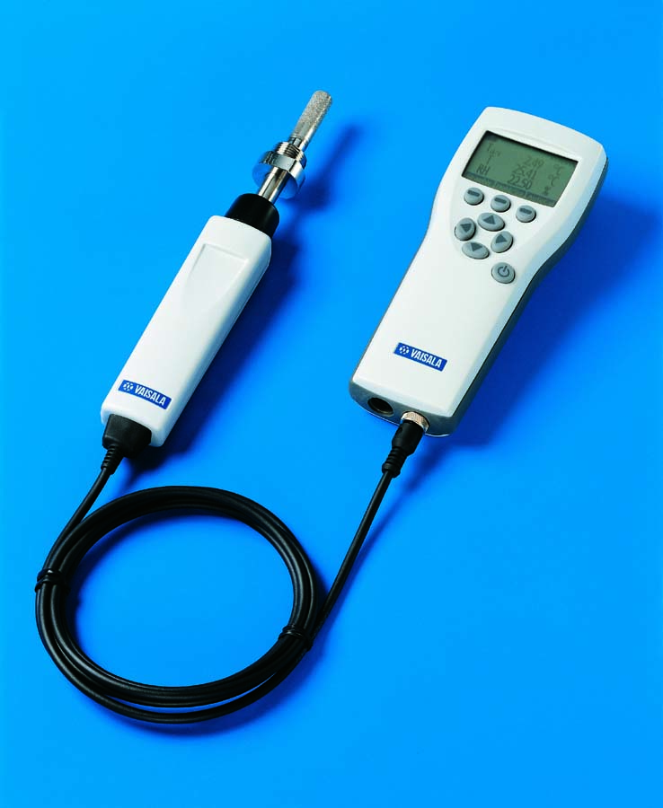
When discussing a typical facility’s compressed air system, it’s helpful to divide the entire network into two separate subsystems: the supply side and demand side. The supply side consists of the compressors and air treatment equipment up to the flow/pressure controller. The demand side consists of the distribution and storage systems or everything after the flow/pressure controller. On the supply side, dewpoint transmitters providing analog signals can be built into the dryer control system or can be installed in-line before or after the receiver tank. On the demand side, fixed mount instruments providing a local display, alarm relays and datalogging capability are quite common throughout the distribution network and before critical end-use applications to give operators and plant personnel a quick assessment of dewpoint conditions at specific points in the system. This helps ensure that the dewpoint level of the air being produced at the dryers is maintained through the entire facility and to the end use points. Portable devices are an excellent tool for verifying dryer performance, conducting quality audits, and checking the calibration of fixed mount instruments.

vaisala monitor

How is dewpoint measured by refrigerated air dryers?
Refrigerated dryers operate by using a refrigerant to cool the supply air with heat exchangers (usually to between 35ºF to 40ºF) and condense out water vapor for removal by a moisture separator and drain. Due to their relatively low initial cost, long term reliability and minimal maintenance requirements, refrigerated dryers often do not integrate a dewpoint transmitter into their design for monitoring or control purposes.

How is dewpoint measured by desiccant air dryers?
Desiccant air dryers can benefit from a dewpoint sensor for monitoring dryer performance, controlling desiccant tower regeneration, or both. Most regenerative desiccant type dryers (heated or heatless) produce a dewpoint of around -40ºC/ºF. Installing a dewpoint instrument with a display or with built in alarm relays to measure the exit air from the dryer is a smart way to ensure good dryer performance. However dryer efficiency can be significantly improved by using a dewpoint device to control the regeneration cycle – known as Dewpoint Demand Switching (DDS). Desiccant dryers operate using two separate towers containing desiccant – one tower is always in operation while the other tower is being regenerated or purged using a portion of the dried exiting air. Some towers switch based on a timer, regardless of whether the desiccant has been fully saturated. By integrating a dewpoint sensor with the dryer control system, the towers will not switch until the dewpoint transmitter senses a degrading dewpoint temperature, thus ensuring full utilization of each desiccant tower and minimizing wasted purge air.

How is dewpoint measured in point-of-use applications?
For point-of-use dewpoint measurements, generally there are two options available, direct in-line insertion or sample extraction. Each method offers advantages and disadvantages that should be considered carefully. Direct insertion involves installing the probe through a threaded connection or “T” in the line. The benefits of this approach are ease of installation with no accessories required and no venting or loss of the compressed air. Line pressure fluctuations and sensor removal however can present drawbacks. The best installation for a dewpoint instrument isolates the sensor from the main line using a stainless steel sample line and sample cell. This setup allows for “valving off” from the main line and the ability to regulate the pressure, which has a considerable affect on the dewpoint reading. Easy installation and removal of the sensor can also be an important advantage.

What are the different prevalent technologies used to measure dewpoint?
With the vast number of different hygrometer technologies currently available on the market for measuring a wide range of dewpoints suited to different applications and industries, it would be difficult to cover all of them here in any detail. I’ll limit the scope of the discussion to briefly address only the most common sensor types used in compressed air measurement.
Condensation hygrometers, often called “chilled mirrors” operate by cooling a surface in a controlled manner until condensation begins to form; this temperature is recorded as the dewpoint temperature of the air. The most common detection method for determining when liquid water has begun to form is optical reflectance, which uses a light source to measure the amount of reflected light from the surface. These devices are well known for their high accuracy (usually +/-0.2ºC dewpoint) but generally require more maintenance to keep their reflective surface clean. They become prohibitively expensive for measuring very low dewpoint temperatures.
Aluminum oxide, silicon oxide, and various other capacitive sensors share some common traits. In all cases, a capacitor is formed between two electrodes with a hygroscopic material serving as the dielectric of the capacitor. The hygroscopic material adsorbs or desorbs water vapor in proportion to the amount of water vapor surrounding the sensor. This changes the dielectric constant of the material and therefore the capacitance of the sensor. The choice of dielectric materials is critical to the performance of the sensor. Aluminum oxide is sensitive to very low dewpoints, but peforms less well in atmospheric humidity levels. These sensors can be economical for low dewpoints when compared to chilled mirrors.
Thin film polymer capacitive sensors operate on the same principle as aluminum oxide sensors but use polymers instead of metallic oxides as the dielectric material. Many polymer sensors are optimized for use in atmospheric levels of humidity and are not suitable for the measurement of gases with dewpoint temperatures lower than -20°C. However, some polymer sensors are designed for low dewpoint measurement, and they typically distinguish themselves by implementing active, automatic self-calibration schemes to monitor and adjust the performance of the sensor. These sensors are cost competitive with aluminum oxide devices and offer the benefit of enhanced long term stability.

What does Vaisala recommend for measurement and monitoring of dewpoint?
When selecting a dewpoint instrument for a particular application, it’s important to consider the following about the installation:
• What is the expected dewpoint level at the intended measurement location?
• What is the pressure range?
• What is the temperature range?
• Will the probe be installed directly in the line or will a sample line be used for external measurement?
• Should the instrument be portable or fixed mount?
• What type of signal output is desired – local display, analog, serial communication?
• What other functionality is of interest – power supply, datalogging, alarm relays?
With this information specified, the field of dewpoint instruments that fit these criteria will be significantly reduced.

A Pharmaceutical Compressed Air System Audit


Pharma audit

A. Introduction

This West Coast pharmaceutical facility has a very clean and organized compressed air system. All equipments is in good working order in the compressor room. The compressor room itself is very clean and well ventilated. The management requested a compressed air system audit for two reasons:
  1. Production problems and downtime resulting from the presence of moisture in the compressed air lines. The compressor room dryers were functioning properly so how could this happen?
  2. Awareness of the high cost of compressed air and a desire to find ways to reduce compressed air demand.
This article will describe the actions taken to address these two issues. The facility operates “24/7” so we have 8,760 operational hours per year. The average electrical rate at this facility is $0.12 kW/h. The power cost formula used is based upon the facility’s current operating conditions of 3.89 CFM/BHP and 95% average motor efficiency.
Power Cost = (BHP *0.746 * 8,760 hours x $0.12 per kW/h) / Avg. Motor Efficiency (95%)
The focus of this audit is on the “Demand Side” with the very top priority being to identify the root cause of the presence of moisture in the compressed air lines.

B. Compressor Room Review

The Compressor Room is extremely clean and well ventilated. There are two rotary screw compressors which are oil lubricated and air-cooled. The air is dried by two parallel refrigerated air dryers. The air then goes into a common header and flows into a 1,040 gallon air storage tank. The air then flows into a Intermediate Flow Controller. From here the compressed air leaves the compressor room and enters the facility. The average CFM per BHP between the two air compressors is 3.80 CFM per BHP.
Compressor Room
Upon entering the compressor room we noted an audible air leak in Compressor #2. We found the leak to be coming from the air end and recommended that the air compressor service provider be contacted immediately. Both air compressors are operating via modulation control. Compressor #2 acts as the base load machine while Compressor #1 is the back-up machine when pressure falls to a predetermined set point.
The two refrigerated air dryers are in good working condition and functioning properly. They are designed to produce a dewpoint range between 33 F and 39 F at a maximum flow of 330 CFM at 100 psi. We took dewpoint measurements at the compressor room outlet (for one week) and found that the average dewpoint achieved was 36 F. This correct dryer performance is what has the facility bewildered by the presence of water in the compressed air lines in the factory. It is worth noting that the dryers are not capable of drying the full air output capacity of the air compressors if factory demand should increase. The dryers have integrated 1 micron particulate filters. We recommend that the facility install a 0.01 ppm oil coalescing filter to protect against oil contamination downstream.
The 1040 gallon air storage tank is adequate for the air demand in the facility. During production we recorded an average air flow of 307.30 CFM which means the tank is providing 3.38 gallons of storage per 1 CFM of air storage. The tank is also piped properly (after the air dryer) with air entering the bottom of the tank and exiting the top – providing more surface area for moisture to be separated and fall to the bottom of the tank.

C. Solving the Problems with Moisture

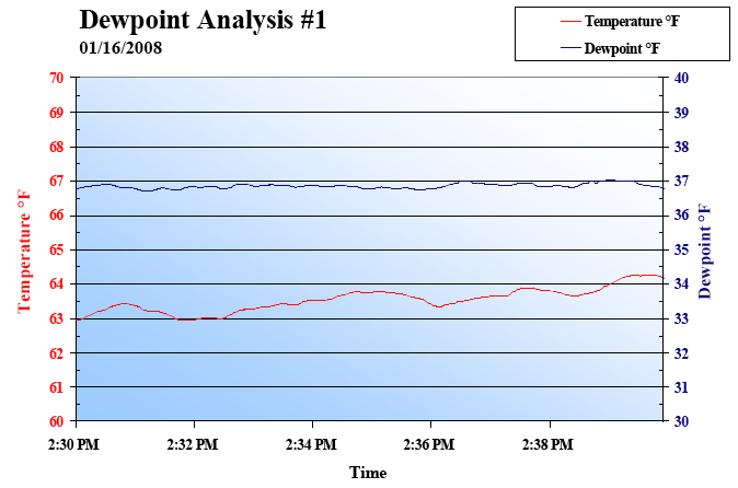
It was initially reported that the plant was “having problems resulting from an excess of water in the compressed airlines”, with the primary area of concern being the small cylinders in the plant. As a standard part of our audit procedure we took dewpoint readings. These test showed a refrigerated drying system according to its specifications. The data depicted below shows a very steady dewpoint with less than half a degree fluctuation over a ten minute time span. The 36 °F average dewpoint is well within the expected range of performance for these dryers.
Dewpoint Analysis
As a result of the ongoing condensation issues, the factory had taken some actions in hopes of remediating the problem. The solution the plant put into place was to install water separators on each line and open the drains on Filter-Regulator-Lubricators (FRL’s) throughout the plant. We found a total of 18 FRL units with the drains open, exhausting approximately 2 CFM each worth of compressed air, totaling 36 CFM worth of compressed air. The problem with this is that the only thing exhausting through the drains was compressed air, thereby decreasing plant pressure at the same time.

Water droplet
Moisture in Pneumatic Cylinders Created Production Down-Time

Adiabatic Expansion

Through further investMoistureigation we found the root of the problem. When air is discharged from the piping between the cylinder and the valve, the temperature of the air drops due to adiabatic expansion. . If the atmospheric dewpoint of the supply air is T1, and the temperature of the air T2 after adiabatic expansion falls below this value (T1
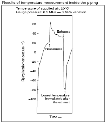
Temp. Measurement

The Solution to Moisture Problems in Pneumatic Cylinders

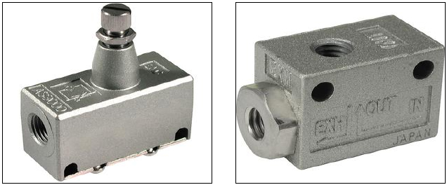
There is a very effective solution to this problem, the installation of quick-exhaust valves directly onto the cylinders. These will allow for the cylinder to fully exhaust on every cycle, thereby eliminating condensation build up and save the cylinders. Along with saving the cylinders in the plant, there is no need to keep the drains of the FRL’s opened in the plant. Leaving the drains opened is actually creating a larger pressure drop throughout the plant, and wasting 36 CFM of compressed air.
Understanding what is happening with the pneumatic cylinders was the key to solving this problem. The facility had been considering installing desiccant air dryers. This would have been a significant capital expense and would not have solved the problem. We were able to identify the adiabatic expansion occurring between the cylinder and the valve. The solution deployed of quick-exhaust valve was a minor expense and achieved with little effort.
Quick Exhaust Valves
Quick Exhaust Valves Solved the Problems with Moisture

D. Demand Side Audit

Aside from solving the downstream moisture problem, our audit reviewed pneumatic circuitry in the facility and also included a compressed air leak audit. Below is a brief summary of some of the opportunities discovered and solved. The end result was that air demand was reduced by 186 CFM. This reduced the plant compressed air demand from an average of 307 CFM to an average of 121 CFM.

The Five Label-Aire Machines

We took note of five Label-Aire machines in the factory. These units were constantly being pressurized even when not being used. We conducted a point-of-use test on these machines and found that each machine consumed an average of 4.2 CFM even when not in use. We only sampled a fragment of time so we are not sure of what percentage of the time the machines are idled but plant personnel tell us that it is a significant percentage of time.
The solution here is to keep the Label-Aire machines from consuming air when idled. This is easily achieved by the installation of two-positioned solenoid valves. The solenoid of these valves will effectively actuate this application only when the product is present. The solenoid of these two-positioned valves can be actuated via several applications such as relay sensors and electronic signals. The table below shows the savings opportunity.

Label-Aire Machines Air Consumption Costs When Idle

Table 2
The Ten Blow Guns
There are ten blow guns at the facility which use inefficient nozzles. This can be costly in the long run due to the decreased impact pressure and increased waste of compressed air. We recommend using high-efficiency nozzles, which can reduce air consumption by 50-75% while increasing the impact pressure at the work surface. These high-efficiency nozzles utilize the Venturi effect to gain efficiencies.

Nozzles
High Efficiency Nozzles on Blow Guns
The Cutter Machine Causes Plant Over-pressurization
The cutter machine is causing the entire plant to run at 100 psig because it requires 95 psig. We conducted a point-of-use test on this machine to verify its’ air usage. The machine was operating between 85 and 90 psig when we started the test. Actual pressure changed over time from 71 psig to 104 psig with 90 being the average. Air flow went from 20 cfm to 10.4 cfm with 4.3 cfm being the average. The cutter was actuating twice a minute, in one minute and 15 second cycles with an intermitten down period of approximately two and one-half minutes.
Due to the intermittent demand of this application, we are recommending the use of a pneumatic booster coupled with a air receiver. This will allow us to reduce the air pressure across the entire facility.
Air Leaks
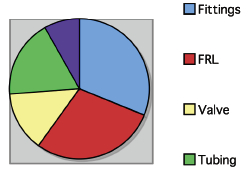
Chart 1
Leak locations in % at the facility 
We have identified and tagged 28 compressed air leaks in the facility. They account for 120 CFM equating to 39% of the plants’ average air flow of 307 CFM. Leaks were found on the airend of one air compressor, and plant-wide on FRL’s, fittings, gauges, and pneumatic tubing.
It is important to have a leak remediation campaign in place to keep leaks from consuming unnecessary compressed air. A proactive approach to leak detection should include all individuals within the plant and the education of machine operators on the cost of leaks.
Machine operators could then combat leaks as they develop by immediately tagging them and notifying maintenance. This approach to leak remediation is perfect for leaks that can be easily felt and heard. Some leaks cannot, unfortunately, be detected by the human ear. We utilize ultrasonic leak detectors to find leaks that are out of range and hard to hear or feel.

E. Capture the Savings

Air Leak
Air Leak on FRL
Multiple holes
Multiple Holes/Leaks in Tubing
Air Leak
Air Leak on Hose/Fitting Connect
The demand side audit made it possible for us to reduce average air demand from 307 CFM to 121 CFM. We fixed leaks (120 cfm), closed open drain valves (36 cfm), and will reduce over-all plant pressure (30 cfm). We now need to look at how the compressors are operating and if they controls are set to capitalize on the new compressed air demand profile.
The “Before” situation was this to produce 307.3 cfm:
  1. Compressor #1 ran 100% loaded in modulation mode. At 100% power it had 90 BHP which equated to $74,292 in annual costs of operation.
  2. Compressor #2 ran 20% loaded in modulation mode. At 78% of power it consumed 67.3 BHP equating to $11,106 in annual energy costs of operation.
  3. Total annual energy cost of operation was $85,398
The “After” situation was this to produce 121.3 cfm:
  1. Compressor #1 was placed on standby for emergency situations. $0 energy cost.
  2. Compressor #2 was capable of having its’ controls modified to Load/No-Load. We ran the machine 37% loaded to meet the demand. At 55% of Power it had 47.45 BHP equating to $39,168 in annual energy costs of operation.
  3. Total new annual energy cost of operation was $39,168
The new annual energy costs to run the air compressors represents a savings of $46,230 per year.
Conclusion
Understanding pneumatics is core to conducting a strong demand side audit. Pneumatic circuits were where the audit was able to discover the dewpoint problem and the opportunities to reduce air consumption and pressure. Understanding air compressors and air compressor controls then allowed the installations’ energy costs to be reduced as a result of the demand side improvements.

Air Quality in the Pharmaceutical Industry


The United States accounts for roughly half of the global pharmaceutical market. This certainly keeps the Food and Drug Administration (FDA) busy in its oversight of pharmaceutical safety and effectiveness, including with the production processes. As the pharmaceutical industry has grown, so too has its utilization of compressed air for breathing air, operation of equipment and instrument air.
PillsThe FDA has taken notice, of course, and the quality of the air being used is a concern; and, rightly so, no standard has been issued for the use of compressed air in production.
“A one-size-fits-all standard won’t work here,” says Dr. Ed Golla, Laboratory Director for Austin, Texas-based TRI Air Testing. “Everyone is doing something a little different.”
While this leaves pharmaceutical companies a somewhat in the dark as to how to vet and confirm the quality of air being used, it does not mean companies need to find the tightest standard possible. Each facility will have unique needs, and the standard applied should best serve these needs.

UNDERSTANDING YOUR FACILITY

In the absence of specified standards governing compressed air quality testing in the manufacturing process or production of pharmaceutical, medical device, and food applications, it is often best to use composite, site-specific testing programs. This may be the most assured way to produce valid, repeatable testing results that will reinforce your site’s quality. Direct Product Contact, Indirect Product Contact, USP and ISO 8573 air standards are common sources from which to draw. A routine testing schedule for your compressed air quality program should provide the appropriate verification and compliance each facility will need for OSHA, FDA and cGMP.
It is imperative that you understand the real needs of each site. You do not need to expend the time and money to establish a quality of air that you don’t actually need. “Clean room” air in a non-clean room air operation, for example, is unnecessary. Specifying air to that level requires expensive equipment to clean and maintain it.
If you bring clean room-level air into a non-clean room environment the quality of the clean air is decreased to the level of the room environment. You’ve brought a small amount of clean air into a larger volume of air at a lower level.
“What they really need to do,” says Dr. Golla, “is first look at the quality of air as it is now--as you’ve been using it for the last 10 or 15 years. Identify that quality and base your spec on the quality of air you have input from the engineers involved in the actual process you’re using.”

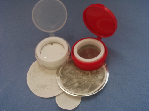
Filters Used

Greater operational efficiency in air management and cost control can be found in understanding the true air needs in each area of your facility rather than using a single approach for the whole facility.

TRI Air Testing works with many pharmaceutical companies in helping them assess and identify the right air quality tests for compliance, operational efficiency, and safety. TRI sends out its testing equipment along with appropriate sampling media. Clients return the samples for analysis and reports are available within 24 hours.
“The depth and stringency of ISO 8573 may be perfectly applicable to an operation’s clean room particulate control,” says Dr. Golla, “but it isn’t right for all applications.”
Facilities with clean room needs, such as those handling implantable devises (e.g., knee and hip joints, defibrillators and pacemakers) cannot have particulate matter on their surface and affecting the safety of product. These facilities must utilize a spec that ensures a heightened level of particle control.
“You can’t use a spec allowing 5 mg per cubic meter or 1 mg per cubic meter or even a 0.1 mg per cubic meter,” says Dr. Golla. “You need information about the individual particle sizes and counts. With implantable devises once you clean the surface and blow it with compressed air you don’t want to leave particles on it.”
The ISO 8573 spec Class 1 or Class 2 requirements may be ideal here.
But for operations that might be using compressed air to blow out bottles before putting in tablets or simply running a piece of machinery, that most stringent air standard may be excessive. It may lead to greater operational expense or present the facility with a test that is difficult, if not impossible to pass—and all on a process that does not actually need that high of a quality of air.
“If they are concerned about particles, but do not necessarily need something like 8573 Class 1 or Class 2,” says Dr. Golla, “they can use a point-of-use filter.”
If only 1% of a facility, for example, uses a very high-quality air for a specific application, but the general air use in the rest of the facility does not need that quality of air, a point-of-use filter may be the right solution and help achieve greater operational efficiency.
One recommendation: engage the production engineers who are most familiar with the air quality needs. Ask them if there is anything that shouldn’t be in the air. Does it need to be filtered to a finer level? Does the air need to be moist? Or does it need to be dry?
Understanding the real impact of air quality on the specific zone of work, the people conducting the work, and the product being produced or serviced will go a long way towards guaranteeing compliance, levels of safety and the quality the FDA and other regulatory entities want to see.

THE CHALLENGE OF MOISTURE

Identifying a proper moisture spec is not easy. Many facilities just use refrigerated air. They cool the air to remove water; then, they bring it back to room temperature so the humidity is relatively low and you won’t get water formation and bacteria growth.
“That’s probably what you need in 90% or more of the operations,” Dr. Golla says, “yet, we put out a guideline to say there ought to be at least a -10°F for indirect product contact and -50°F or so for direct product contact. I think we put a pretty big asterisk on that to recognize that this one may or may not apply.”
Certain products will be destroyed by -50°F degree dew point air. It will desiccate and deactivate them. For other products, the moisture could react with the material and cause problems, so you might need extremely dry air.
Just as ISO 8573’s most stringent requirement classes are not right for all particulate testing of compressed air, there is no single approach to dew point and moisture management.

Air Filtration in the Pharmaceutical Industry, Cleanroom Filtration


Sterile FillThe manufacture of pharmaceutical, bio-pharmaceutical and medicinal products is unique from many other manufacturing processes for a number of reasons.  The most important is that these products are utilized for the health and well being of living organisms, including humans.  Because of this, the design of the manufacturing area of these facilities is required to follow Good Manufacturing Practice (GMP).
The US Food and Drug Administration (FDA) has published Federal Regulation 21 CFR Part 820, Quality System Regulation.  This regulation follows ISO 9000.  More specific to the clean areas and cleanrooms of interest are the standards and drafts under ISO 14644.   A good overview of these standards is available on the IEST web site.  Similarly, European Standards provide European Nations with guidelines for GMPs.
Why do these GMPs matter with regard to air filtration in pharmaceutical, bio-pharmaceutical, and medicinal products manufacturing processes?  Because GMPs require that the quality of these products be assured, including the control of product contamination during manufacturing and packaging.  Generally speaking, the control of contamination is most critical for injectable or parenteral preparations as compared with topically applied or orally ingested preparations.  Eye drops, which can be considered a topically applied preparation, are an exception to this rule as they eye is quit susceptible to infection.

Wednesday, August 21, 2013

Pharmaceutical Sterility Testing


Essential things to know


By Steven Richter


Sterility testing of pharmaceutical articles is required during the sterilization validation process as well as for routine release testing. USP1 requirements employ sterility testing as an official test to determine suitability of a lot. An understanding of sterility testing is beneficial in terms of designing a validation process. The need to provide adequate and reliable sterility test data is an important quality assurance issue. Sterility testing is a very tedious and artful process that must be performed by trained and qualified laboratory personnel. The investigation of sterility test failures is a process that requires attention to environmental data as well as many other factors including training and sample difficulty.

This paper presents the general concepts and problems associated with sterility testing as well as the various testing methodologies. Most USP <71> sections are harmonized with the EP/JP.

Sterility testing is an essential part of every sterilization validation. Sterility testing is an extremely difficult process that must be designed and executed so as to eliminate false positive results. False positive results are generally due to laboratory contamination from the testing environment or technician error. The testing environment must be designed to meet the requirements of the United States Pharmacopeia (USP) in terms of viable microbial air and surface counts. Growth media used in sterility testing must be meticulously prepared and tested to ensure its ability to support microbial growth. Procedures for sampling, testing, and follow-up must be defined in the validation procedures.

Sampling Plans


The official test, the USP (Volume 30) recommends testing 40 units per production lot. A reprint of Table 2 "Minimum Quantity to be Used for Each Medium2" is on the next page. Some of the quantities are not harmonized with the EP/JP volumes.3

For combination products, the ISO 11137/111354 standards recommend various sterilization validation sampling plans based on lot size and validation method. In cases where small lots (>1000) are manufactured, the sampling size depends on lot size.

Environmental Concerns Related to Sterility Testing


The sterility test environment is described in USP General Informational Chapter <1211>. The environment should be as stringently controlled as an aseptic processing environment. An aseptic processing environment (clean room) is used to dispense sterile pharmaceuticals into presterilized containers. A clean room is generally a room that delivers laminar flow air which has been filtered through microbial retentive High Efficiency Particulate Air (HEPA) filters. The room is maintained under positive pressure and has specifications for room air changes per hour. An environment used for sterility testing should be similar in design to an aseptic processing environment; there should be an anteroom for gowning and a separate area for the actual sterility testing. The testing area should meet ISO Class 5 particulate control requirements (specified in USP chapter (1116)). Sterility testing should not be carried out under a laminar flow hood located within a room that is not maintained as ISO Class 5. Along with particulate testing in the environment, the laboratory must test for viable bacterial and fungal organisms ubiquitous to it. The sterility test technician must be suitably gowned in sterile garments that prevent microbial shedding into the room. The room should be validated in terms of particulate and microbial levels. The laboratory must have a validation and training program for gowning and sterility testing.

Our validation programs require that technicians consecutively test 40 simulated samples for both membrane filtration and direct immersion methods without a false positive test result under less than ideal environmental conditions. Isolator technology is utilized to create a sterile environment for one to test pharmaceutical articles. The validation required to qualify an isolator is extensive. The isolators are generally sterilized using chemical sterilization.

Many issues surround the robustness of the sterilization process. Qualifying and maintaining an isolator system for sterility testing may require extensive work. In testing pharmaceutical articles in a closed system such as SteritestTM, an isolator may not be the best cost approach to the environmental concerns. Most environmental concerns can be obviated by standard aseptic processing GMP's.5

Methodologies


The United States Pharmacopeia is a compilation of validated methods and official monographs for pharmaceuticals and medical devices. IT is broken down into the following sections: Monographs, General Informational Chapters, and General Requirements. General Informational Chapters <1000> series are not legal requirements. The Sterility Test (USP Section <71>) is categorized under General Requirements and is therefore a legal requirement.

For combination products, the ISO radiation sterilization microbial methods (11737-2 1998)6 describes a sterility test which is a modification for the USP method. This test is specific for the detection of aerobic organisms that have been exposed to sub-lethal sterilization cycles. This ISO sterility test method is recommended for the validation of both gamma and electron beam sterilization processes.

The method of choice for EO7 sterilized products is the official USP <71> procedure.

Processes


Prior to actual sterility testing, it is prudent to send an example sample to the testing laboratory so the laboratory can determine the appropriate testing procedure. Each product should have a unique procedural specification for testing. The procedure should be very specific in terms of which items (or vials/syringes) to test. The procedure must indicate the Sample Item Portion (SIP). The Sample Item Portion is the percentage of the complete product tested. Since medical devices come in all shapes and sizes, it is very difficult to test large and cumbersome medical devices in their entirety. Therefore, the test laboratory will determine a Sample Item Portion which is a portion of the sample expressed in fractional terms (i.e. 0.1 for 10% of the sample).

This number is used in gamma and electron beam dose setting methods. The SIP portion should be validated by sterility testing.

Combination products have unique challenges. A combination product is defined as one that has a drug component with medical device. For example, a drug coated stent. The agency's Office of Combination Products (OCP) would determine which regulatory branch (CDRH, CDER or CBER) is officiating the product. Official USP sterility testing of combination products is required for all sterile drug products. The drug product component applied aseptically creates the largest challenge to laboratory personnel. Biologics must be aseptically processed and cannot be terminally sterilized. In the near future, we will see more biologics that are combination products. Combination products sterilized by radiation are generally handled as medical devices following the ISO 11137 standard. For the most part, pharmaceutical GMPs would take precedent over 820 QSR8 requirements with all combination products. The more robust GMP9 requirement would assure reduced bioburden counts and consistent microbial populations during manufacturing.

The USP <71> Sterility Test contains two qualifying assays which must be performed prior to sterility testing. They are the "Suitability Test" (Growth Promotion Test) and the "Validation Test" (Bacteriostasis and Fungistasis Test).

The Suitability Test is used to confirm that each lot of growth media used in the sterility test procedure will support the growth of fewer than 100 viable microorganisms. If the media cannot support the growth of the indicator organisms, then the test fails. Secondly, a portion of each media lot must be incubated and assessed for sterility according to the incubation parameters (time, temperature) established by the method. If the media is found to be non-sterile, then the test fails.

The Validation Test is used to determine if the test sample will inhibit the growth of microorganisms in the test media. Stasis, in terms of microbiology, is defined as the inability of a microorganism to grow and proliferate in microbiological media. Media that is bacteriostatic does not necessarily kill bacteria; it simply may retard bacterial growth and proliferation. The Validation Test must be performed on each product prior to and/or during sterility testing. This test determines if the media volumes are valid for the particular product. Some medical products contain bacteriostatic and fungistatic compounds that may require special procedures and special media for testing. This test is similar to the Suitability Test described above, however, the product sample is placed in the media along with the microorganisms. Microbial growth in the presence of the test samples is compared to controls without test samples. If microbial growth is present in the sample and control containers, then the test is valid. The next step is to proceed to actual sterility testing. Suitability, validation and sterility tests can be performed simultaneously.

The USP describes three general methods for sterility testing: 1) Membrane Filtration, 2) Direct Transfer (Product Immersion); and 3) Product Flush.

Membrane Filtration Sterility Testing


The Membrane Filtration Sterility Test is the method of choice for pharmaceutical products. It is not the method of choice for medical devices; the FDA may question the rationale behind using the membrane filtration test over the direct transfer test for devices. An appropriate use of this test is for devices that contain a preservative and are bacteriostatic and/or fungistatic under the direct transfer method. With membrane filtration, the concept is that the microorganisms will collect onto the surface of a 0.45 micron pore size filter. This filter is segmented and transferred to appropriate media. The test media are fluid thioglycollate medium (FTM) and soybean casein digest medium (SCDM). FTM is selected based upon its ability to support the growth of anaerobic and aerobic microorganisms. SCDM is selected based upon its ability to support a wide range of aerobic bacteria and fungi (i.e. yeasts and molds). The incubation time is 14 days. Since there are many manipulations required for membrane filtration medical device sterility testing, the propensity for laboratory contamination is high. Therefore, in an open system, more sterility failures are expected when using this method. A closed system is recommended for drugs and small devices or combination products. Most pharmaceutical articles are tested using a closed system. In closed systems, the propensity for extrinsic contamination is very low.

Direct Transfer Sterility Testing


Combination products: This method is the method of choice for medical devices because the device is in direct contact with test media throughout the incubation period. Viable microorganisms that may be in or on a product after faulty/inadequate sterilization have an ideal environment within which to grow and proliferate. This is especially true with damaged microorganisms where the damage is due to a sub-lethal sterilization process. All microorganisms have biological repair mechanisms that can take advantage of environmental conditions conducive to growth. The direct transfer method benefits these damaged microorganisms. The entire product should be immersed in test fluid. With large devices, patient contact areas should be immersed. Large catheters can be syringe filled with test media prior to immersion. Cutting catheter samples to allow for complete immersion is the method of choice.

The USP authors understand that appropriate modifications are required due to the size and shape of the test samples. The method requires that the product be transferred to separate containers of both FTM and SCDM. The product is aseptically cut, or transferred whole, into the media containers. The test article should be completely immersed in the test media. The USP limits the media volume to 2500 ml. After transferring, the samples are incubated for 14 days.

Product Flush Sterility Testing


Combination products: The product flush sterility test is reserved for products that have hollow tubes such as transfusion and infusion assemblies where immersion is impractical and where the fluid pathway is labeled as sterile. This method is easy to perform and requires a modification of the FTM media for small lumen devices. The products are flushed with fluid D and the eluate is membrane filtered and placed into FTM and SCDM. This method is not generally used.

Bulk Drug Products / Biologics and Pharmaceuticals


Bulk Pharmaceuticals (APIs) are tested for sterility per USP 71 prior to release to the manufacturing processes.

Bulk Biologics are tested according to 21 CFR 610.12 for sterility testing. This method requires one media (FTM). The sample test sizes are listed in the document. Volumes are no less than 10 ml.10

Interpretation of Sterility Test Results


The technician must be trained in the method of detecting growth during the incubation period. Growth is determined by viewing the media, which is generally clear and transparent, against a light source. Turbid (cloudy) areas in the media are indicative of microbial growth. Once growth is detected, the suspect vessel is tested to confirm that the turbidity present is due to microorganisms and not due to disintegration of the sample; sometimes samples produce turbidity because of particulate shedding or chemical reactions with the media. Once a suspect container has been tested, it should be returned to the incubator for the remainder of the incubation period. Samples that render the media turbid are transferred on Day 14 of the test and incubated for four days. Growth positive samples require further processing such as identification and storage.

Sterility Test Failure Investigation


For every positive sterility test (OOS), the laboratory should perform an OOS investigation to determine the validity of the positive growth. This investigation encompasses the following items:

  1. clean room environmental test (EER) data;
  2. media sterilization records;
  3. technician training records;
  4. the relative difficulty of the test procedure;
  5. control data (open and closed media controls);
  6. technician sampling data (microbial counts on gloves and/or garments post testing).

The USP allows for a re-test of the product if persuasive evidence exists to show that the cause of the initial sterility failure was induced by the laboratory. Identification and speciation of the isolate(s) is a significant contributing factor to the final decision. If the First Stage sterility test can be invalidated by the laboratory, then the USP allows for Second Stage sterility testing. Second Stage sterility testing requires double the original number of samples tested. The Second Stage test can be repeated if evidence exists invalidating the test due to a laboratory error as above.

A detailed investigation may uncover circumstantial evidence to support a final decision. It is recommended that sterilization cycle data, environmental data, and bioburden data be reviewed prior to making any decision to release product.

It is recommended that medical device manufacturers qualify the test procedure with non-sterile samples.

The probability of a false positive can be calculated using John Lee's formula.11 The formula is based upon sample container diameter, amount of time container is left open and the room particulate count.

Sterility testing requires high levels of control with regards to GMPs, Good Laboratory Practices12, environment (aseptic clean room ISO class 5 or better), and employee practices. It is essential that meticulous technique be employed in the practice of sterility testing. Sterility testing is an integral part of sterilization validation as well as a routine quality control. Generally, false positive results are uncommon in testing drug products using a closed system. Combination products have challenges that should be planned into a robust QA program.

References


  1. The United States Pharmacopeia, 30th Revision, The United States Pharmacopeial Convention: 2008
  2. USP 30 Table 2 Minimum Quantity to be Used for Each Medium
  3. USP 30 Table 3: Minimum Number of Articles to be Tested in Relation to the Number of Articles in the Batch
  4. ISO 11137 Sterilization of health care products – Radiation – Part 2 2006: Establishing the sterilization dose
  5. FDA Guidelines 2004 "Guidance for Industry Sterile Drug Products by Aseptic Processing, Current Good Manufacturing Practices," September, 2004
  6. ISO 11737 ANSI/AAMI/ISO 11737-2 1998 – Sterilization of Medical Devices – Microbiological Methods – Part 2, Tests of Sterility Performed in the Validation of a Sterilization Process
  7. ISO 11135 1994 Medical Devices Validation and Routine Control of Ethylene Oxide Sterilization
  8. Code of Federal Regulations Title 21/Chapter I/Part 820, "Quality Systems Requirements: General," 2006
  9. GMPs CFR 201 Title 21 2006
  10. 21 CFR Part 610.12 Bulk Biologics
  11. Lee, John Y. "Investigation Sterility Test Failures" Pharmaceutical Technology, February 1990
  12. Code of Federal Regulations Title 21/Chapter I/Part 58, "Good Laboratory Practice for Nonclinical Laboratory Studies," 2006

FDA STERILITY DRAFT GUIDE RECOMMENDS "STERILE MEDIA FILL" TECHNIQUE TO VALIDATE ASEPTIC ASSEMBLY PROCESS; VALIDATION, LAB CONTROLS, STERILITY TESTS ADDRESSED

Executive Summary

FDA's newly released draft guideline on sterile drug products produced by aseptic processing recommends the use of "sterile media fills" to validate the assembly process. The guideline defines "sterile media fills" as microbiological growth nutrient media used to simulate sterile product filling operations. According to the draft guide, "media filling in conjunction with comprehensive environmental monitoring can be particularly valuable in validating the aseptic processing of sterile solutions and suspension." In general, the guide notes, validating the aseptic assembly of sterile product elements, i.e., filling/sealing, has generated more questions from industry than has the sterilization of those elements, and "therefore, the guidance presented places greater emphasis on validating the aseptic assembly operations." Questions raised by industry about media fills and addressed in the guideline concern contaminating equipment with media, frequency and number of runs, size of runs, the media itself, environmental conditions, and test results. FDA considers the aseptic processing guide to be a priority guideline due to the frequency of compliance problems in the area, the degree of unfamiliarity in the industry with FDA expectations, and the lack of uniformity between firms in their sterile practices. The agency explains that the guideline is not intended to cover all aspects of sterile processing but to address those areas that have been most problematic for industry. Selected current good manufacturing practice requirements are cited, followed by a discussion of practices and procedures which FDA considers as acceptable means of meeting the requirement. The guide discusses buildings and facilities, components, containers/closures, production time limitations, validation, laboratory controls, and sterility testing. Guide Stresses Importance Of Specifying Criteria For Conducting Investigations Of Sterility Failure Another aspect of aseptic processing that the guideline addresses at some length is drug product sterility testing. Aspects of the sterility testing which the guideline states "are of particular importance" and discusses include control of the testing environment, understanding the test limitations, interpretation of positive results, and retesting. The guideline emphasizes the importance of having written procedures in place to specify the criteria for conducting investigations of sterility failures, including decision rules for rejecting batches. Firms should lean toward the side of safety and reject batches if the investigation of positive testing results shows a production failure. A retest is warranted, the guide states, only if each of three conditions is met: "The organism found in the test is not found in the production area or the bioburden, but has been shown to occur in the testing area; the incidence of growth in test units is not greater than the laboratory's record of false positives over a period of time; and batch production and control records show no discrepancies which could affect sterility." Aspects of asceptic processing not addressed in the draft document include employee hygiene, aseptic gowning, and clean room design. However, FDA said that "these and other apsects will be covered in future revisions of this guideline as needed." The guideline also does not address terminally sterilized drug products, although some portions may be applicable to their preparation, FDA noted. The Feb. 1 Federal Register notice of availability of the draft guide requests that comments be submitted by May 2, 1985. The draft is available from, and comments should be submitted to the Dockets management Branch, FDA, Rm. 4-62, 5600 Fishers Lane, Rockville, MD 20857.

Media Fill Validation Test in Sterile Pharmaceutical


Procedure for aseptic filling or media fill validation in pharmaceuticals, frequency, number of runs and interpretation of results.


PREREQUISITES

1. Approved Soybean casein digest broth.
2. Aseptic area gowning procedures and Entry into sterile areas.
3. Environmental Monitoring of manufacturing areas by Plate Exposure, Air sampling and surface monitoring procedures and its SOP’s. Personnel Monitoring by Finger Dab & Swab Test Method & its SOPs.
4. Qualified and validated manufacturing equipments, system facility (i.e. HVAC, water, compressed gases) CIP and SIP procedures.
5. Trained operating personnel’s.
6. Approved BMR for media fill trial.

EQUIPMENT / SYSTEM DESCRIPTION:

Location : Manufacturing Area ( Mixing room and filling room)
Equipments : Mixing Tank, Holding Tank, Filtration housings, connected product line and FFS machines

IDENTIFICATION OF CRITICAL CONTROL MONITORING PARAMETER

Critical control parameters were identified and it should be considered and recorded during validation program, following are the critical points-
• Check all the equipment and system facility is validated.
• Check and ensure that the HVAC system, compressed air, CIP and SIP procedures are qualified.
• Check and ensure that all operations, cleaning/ sanitization procedures are established and operating personnel are trained.
• Check the Media used for Process Simulation is passed for GPT
• Check and ensure that the WFI used for preparation of batch is complied to USP/IP
Environmental monitoring should cover three operational shifts, by following methods –
• Settle plate method
• Air sampling
• Swab testing
• And Personnel monitoring

STUDY DESIGN

Worst Case Consideration:

1 Allowed maximum number of personnel in the Aseptic Processing Area, including the maintenance and housekeeping personnel
2 Simulating routine machine parts assembling / disassembling, equipment / system setups, in between minor maintenance jobs
3 Increased the time period to start the filling operation
4 Duration of the media fill trial was more than that required for routine manufacturing operation.
5 Simulating Process / Power breakdown during the process simulation test
6 Shift changes and breaks

Frequency, Duration and Number of runs:

1 Media fill trials must be performed on semi-annual basis for each aseptic process and additional media fill trials should be performed in case of any change in procedure, practices or equipment configuration.
2 Filled units in Media Fill run should be 10,000 units or more. Fill minimum 3000 units in each production shift.
3 The duration of Media Fill run must cover all the three operational shifts in each run turn by turn including worst cases.
4 Fill volume for Media Fill run for LVP is 60 ml..

Environmental Consideration:

1 Cleaning of Area must be done by using routine cleaning agent and disinfectant solution, as per latest SOP
2 Microbiological Environmental monitoring should be carried out to cover the entire media fill program for manufacturing area by Settle plate, Active Air sampling, Swab test and personnel monitoring as per the latest SOP.

Media:

1 Soybean Casein digest Medium, manufactured by Hi Media Laboratories should be used for Media fill trial.
2 The media must be passed the test for GPT to promote the growth of gram-negative and gram-positive bacteria and yeast and molds.

Incubation and examination of filled units:

1 Incubate all media filled units in normal position after leak test at of 20 to 25deg.C for 7 days. Incubation temperature should be maintained within 22.5 ± 2.5deg.C.
2 After completion of 7 days Incubation at 20 to 25deg.C, invert the units and incubate them at 30-35deg.C for next 7 days. Incubation temperature should be maintained within 32.5±2.5deg.C .
3 Each media filled unit should be examined by trained Microbiologist after 3rd day, 7th day, 10th day and 14th day.
4 All suspect units identified during the observation should be brought to the immediate attention of the QC Microbiologist.

Interpretation of Test Result:

1 Any contaminated unit should be considered objectionable and investigated. The microorganism should be identified to species level.
2 The investigation should survey the possible causes of contamination.
3 When filled units up to 10000, one contaminated unit should result in an investigation, including consideration of a repeat media fill.

VALIDATION PROCEDURE

CIP and SIP for LVP line:

1 Carry out cleaning of LVP mixing tank and holding tank along with product line and bottle pack machine 360 as per SOP for CIP.
2 At the end of cleaning, collect last rinses sample from sampling point and send to QC department with written information for testing of previous product traces.
3 After getting approval report from QC, affix status label on the tank “READY FOR STERILIZATION”.
4 Immediately carry out the sterilization of LVP holding tank along with final filter and product line of bottle pack machine as per its respective SOP.
5 After Sterilization, affix a status label on the LVP line.

Dispensing of Soybean Casein Digest Medium for 750 L batch size:

1 Give raw material requisition slip in duplicate to store (either computerized or manual) duly signed by Production Officer and Authorized by HOD (Production)
2 Enter to dispensing room as per SOP for entry exit procedure to dispensing area.
3 Check for the clearance of the area from any unwanted materials. Check for the cleanliness of the area, LAF, weighing pan as per checklist. Put “ON” the reverse LAF unit 15 minutes before dispensing of material.
4 Check the availability of clean containers, SS scoops, pressure differentials, and temperature & humidity should be not more than 25deg.C and 45 to 60% RH respectively.
5 Check the balance spirit level is within the circle and then calibrate the balance as per Balance Calibration SOP.
6 Take the Approved Soybean Casein Digest Medium in pre-dispensing room, place on SS pallet and check the label of container for correctness and Approval of material.
7 Transfer the material to Dispensing room, place the empty clean container on the balance and record the tare weight. Press “ZERO” of the balance and weigh the required quantity of material, note the weighed material and then remove the container from balance and press Zero.
8 Close the dispensed material, affix the weighing tag and transfer the material in dispensed material storage room.
9 After dispensing, put “OFF” the balance and LAF. Clean the surrounding area, balance and spray with 70% IPA solution.
10 Reseal the original container and shift to their original place.

Batch Preparation 750 L:

1 Ensure that the area and product line is clean and free from the traces of previous product.
2 Recheck the tag and gross weight of Soybean casein digest medium (SCDM) to be used for manufacturing and ensure that they match as per entries made in the BMR weighing sheet.
3 Check the status board affixed on the tank “READY FOR USE”, also verify the records and ensure that the bottom outlet valve of the mixing tank is closed.
4 Send the entry point sample of WFI from the user point to QC department for testing along with BMR.
5 On approval of WFI sample from QC department, affix a status board on the Mixing tank “UNDER MANUFCTURING” with Product name and B. No.
6 Collect approx 200 L water for injection at 80 to 85deg.C in a manufacturing tank fitted with stirrer.
7 Start the stirrer and add SCDM through the main hole of the tank.
8 Continue stirrer for complete dissolution of ingredients.
9 Stop the stirrer.
10 Make up the volume to the 750 L with water for injection.
11 Start the stirring for complete dissolution of SCDM and homogeneous bulk solution (generally required 10 minutes).
12 Collect sample of bulk solution in a sterile sampling bottle and send it to QC for testing of color clarity, pH and bioburden along with bulk intimation slip.
13 After getting clearance of bulk analysis from Quality Control, start the filtration from mixing tank to Holding tank with the help of pump as per its respective SOP.
14 Perform the bubble point test of the final filter after holding tank as per Bubble point test SOP.

Filling And Sealing:

1 Start the filtration from holding tank to FFS machine using pump.
2 Drain one buffer tank approx 1.3 liters of bulk solution from filling nozzle to eliminate any possibility of dilution of bulk by condensates in product line of the machine post SIP
3 Check online cartridge filter integrity test.
4 Start Machine and discard initial 10 shots.
5 Collect first cassette of vials from next shot and send the sample with written information to QC for testing.
6 Arrange the out coming cassettes of vials sequentially in vacuum chamber tray and verify the results of testing from QC department.
7 Now start the filling and sealing continuously as per SOP for Filling and sealing.
8 Collect the filled and sealed containers coming out of the filling area in plastic crates.
9 During filling operation keep the filled vials separately for each breakdown, shift change, power breakdown, stoppage etc and assign lot number.
10 Carry out the leak test.
11 After leak test, transfer the goods vials in the clean plastic crates horizontally in cassette from one above the other, lot wise separately.

Incubation and Examination of Media Filled Units:

1 Incubate all media filled units in normal position after leak test at 20 to 25deg.C for 7 days. Incubation temperature should be maintained within 22.5 ± 2.5deg.C .
2 After completion of 7 days Incubation at 20 to 25deg.C, invert the units and incubate them at 30-35deg.C for next 7 days. Incubation temperature should be maintained within 32.5±2.5deg.C .
3 Each media filled unit should be examined by trained Microbiologist after 3rd day, 7th day, 10th day and 14th day.
4 All suspect units identified during the observation should be brought to the immediate attention of the QC Microbiologist.
5 During incubation, if any unit found to be damaged should be recorded in media fill observation format.

Interpretation of Results:

1 When filling fewer than 5,000 units, no contaminated units should be detected.
2 When filling 5,000 to 1,0000 units :
a. One contaminated unit should result in an investigation, including consideration of a repeat media fill
b. Two contaminated units are considered cause for revalidation, following investigation.
3 When filling more than 10,000 units :
a. One contaminated unit should result in an investigation;
b. Two contaminated units are considered cause for revalidation, following investigation.

Failure Investigation:

1 Any contaminated unit should be considered objectionable and investigated. The microorganism should be identified to species level.
2 The investigation should survey the possible causes of contamination during media fill trials i.e. Environmental, personnel and surface monitoring.
3 Based on the outcome of the investigation, assign the cause of failure is assignable or not assignable.
4 If the cause is assignable, then take a corrective and preventive action and record the same in suitable format.
5 If the cause is not assignable, then the process should be validated, as it is a new process. Consecutive three-process simulation test should be performed to demonstrate consistency and reliability on the sterile formulation manufacturing process to produce acceptable product.


Process simulation

Process simulation studies should be designed to emulate the routine production process as closely as possible, including formulation, filtration and filling stages. Processes will vary in relation to the type of product to be filled, e.g. liquid or solid dosage forms, and each process simulation is a unique event whereby extrapolation of outcomes cannot be directly linked to actual process contamination rates.

The study will be performed using microbiological growth media in place of active pharmaceutical ingredients (API). This is a 'worst case' senario as most pharmaceutical products normally would not support microbiological growth. The selection of the medium should be based on its ability to integrate into the process at the earliest formulation stage and therefore have the capacity to be introduced to the filling process by filtration. Also the growth promotion characteristics should allow recovery of the typical flora recovered from environmental monitoring programs. The microbiological culture media itself can potentially be a source of contamination so to avoid a culture media related positive fill test, the media is irradiated and can be presented either in the dehydated format or as a ready to use broth.

Modern culture media, designed for media fill trials, possess certain attributes that facilitate process simulations; they will be irradiated making them suitable for introduction into compounding areas, will dissolve in cold water and have known filtration performance as standard broth can be slow to filter or block the filter. Also, those who wish to use an animal-free product can now obtain a vegetable alternative.

Following formulation, filtration and filling the closed vessels are incubated and inspected for contamination.

VACCINE AND SERUM EVILS


BY
HERBERT M. SHELTON

Vaccinia

Vaccinia is an acute infectious disease caused by vaccination. Vaccination is the inoculation of child or adult, well or sick, with septic matter (pus) derived from suppurating (festering) sores on the abdomen of a previously infected cow. I think this definition is incomplete in an important respect—I should have said that it is a criminal operation.
The disease dates from about the year 1774 when an ignorant and superstitious English farmer, Benjamin Jesty, vaccinated his wife and three children with matter taken from sores on cows suffering with cow-pox," using a darning needle with which to make the incisions. Jesty believed a superstition, then prevalent among the milk-maids, that, one who had had cowpox was immune to small-pox.
Notes of this daring experiment were made by a doctor Nash who died in 1785. At his death these notes passed into the hands of Mr. Thomas Nash who was acquainted with Edward Jenner, a notorious charlatan, who is credited with having "discovered" vaccination. In 1789 Jenner inoculated his eighteen month’s old son with swine-pox matter. He followed this with other inoculations of other children and the filthy practice of vaccination was definitely launched.
An English writer, Arthur Wollaston Hutton, M. A., says of Jenner’s framing and qualifications: "But his professional acquirements were but slender; his medical degree was the outcome of no examination or scientific work, but merely of a fee of fifteen guineas paid to the University of St. Andrews; while his other and more important distinction, his Fellowship in the Royal Society, was obtained by what even Dr. Norman Moore, his latest biographer and apologist, is constrained to admit was little else than a fraud."
Thus we have a filthy practice, born out of the ignorance and superstition of the past and fathered by an ignorant imposter and fraud, palmed off on the world today as a scientific procedure. It is really remarkable, the number of instances in the history of medicine, of practices and theories now in vogue, that owe their origin to ancient customs, traditions and superstitions.
It is not known how remote was the belief among the cow hands and dairy maids of England in the immunizing potency of cow-pox; but it is thought to have come out of the practice of inoculation which was introduced into England from the East, by Lady Mary Wortley Montague, wife of the British Ambassador to the Ottoman Court, in 1717. The practice was abolished by act of Parliament in 1840, due to its evils. In 1754 the Royal College of Physicians issued the following manifesto, which reads strangely like the statements made by physicians today about vaccination:
‘The College, having been informed that false reports concerning the success of inoculation in England have been published in foreign countries, think proper to declare their sentiments in the following manner, viz.: That the arguments which at the commencement of this practice were urged against it have been refuted by experience, that it is now held by the English in greater esteem, and practiced among them more extensively than ever it was before, and that the college thinks it to be highly salutary to the human race."
Despite this evident lie by this august body, the practice was not successful; it was not highly salutary; and experience did not refute the arguments used against it. It was a very damaging practice which caused an increase in small-pox in England and was finally abolished by law. Edward Jenner, following Benjamin Jesty, grafted the old inoculation practice onto the milk-maid’s creed and vaccination (from vacca—cow) was born.
I mentioned that the inoculation practice was introduced from the east. The date of the origin of this superstitious practice is hidden in the darkness of pre-history. Savage and barbaric peoples, in various parts of the world, practiced inoculation. It is thought to have started in India. where so many of our superstitions originated, and spread from there to Africa and Europe.
From time immemorial the Negroes and Arabs of Nubia practiced inoculation against smallpox. The Ashantees and the Moorish and Arab tribes in Northern Africa practiced arm to arm inoculation from ancient times. Savage tribes of the Upper Congo practiced it to prevent "syphilis." The Baris of Lado inoculated themselves over the left breast. The Negroes in Senegal inoculated their children on the arms. The Moors and Pouls of Senegambia practiced inoculation against pleuro-pneumonia. A practice of this kind was in vogue in Berne, Switzerland in the 18th century.
The first record of smallpox seems to be in India, where also is the first record of inoculation, where the practice was in vogue over three thousand years ago. Dhanwantari, the Vedic father of medicine, and the earliest known Hindu physician, supposed to have lived 1500 B. C., is said to have been the first to practice inoculation and it is also stated that the Hindus employed a vaccine. For over a thousand years inoculation has been practiced in China.
The practice is so mixed up with the religious superstitions of various peoples that its origin may not be difficult for students of religious history to guess. In India, in Malaba and in other sections of the world, inoculation was mixed up with the worship of the smallpox goddess. Inoculation seems to have been nothing more than a superstitious rite designed to placate and appease the wrath of an irascible deity. People who imagined all their sufferings were sent upon them because they had offended some of their gods or goddesses originated the filthy rite to get the goddess into a good humor again.
According to a Mr. Porter, who was English Ambassador at Constantinople in 1755 (Gentleman’s Magazine, Oct. 1755): "It is the tradition and opinion of the country that a certain angel presides over this disease. That it is to bespeak his favor and evidence their confidence that the Georgians take a small portion of variolous matter, and, by means of scarification, introduce it between the thumb and fore finger of a sound person. The operation is supposed to never miss its effect. To secure beyond all uncertainty, the good will of the angel, they hang up scarlet clothes about the bed, that being the favorite color of the celestial inhabitant they wish to propitiate.’
I cannot imagine St. Paul, who refused to eat meat that had been offered up to idols, baring his arm for pus that is being offered up to the goddess of smallpox. I cannot imagine Moses, whose Kosher laws, in most of their essential particulars, are excellent, commanding the Jews to have this trefe stuff inoculated into their bodies.
Symptoms: Vaccinia begins after inoculation with slight irritation at the site of vaccination. On the third or fourth day the eruption appears in the form of a red papule, surrounded by a red areola. On the fifth or sixth day the papule becomes a vesicle, being filled with a watery substance or a clear substance, with a distinct central depression (umbilication). By the eighth day the vesicle is perfected and is then surrounded by a wide reddened zone of inflammatory edema, which is the seat of intense itching. By the tenth day the contents are purulent (pus) and the vesicle has become a pustule. The surrounding skin is now much inflamed and painful. About this time the reddened areola begins to fade and dessication sets in with the gradual formation of a thick brown crust or scab, which becomes detached and falls off about the twenty-first to twenty-fifth day, leaving an ugly scar. The scar is at fist red but gradually becomes paler than the surrounding skin having a punched-out appearance and is pitted. The evolution of this pathology is accompanied with fever and constitutional symptoms, malaise, and enlargement of the adjacent lymph nodes or glands.
Notice the symptoms above described (and this description is gathered from standard medical works) and you will at once realize that we have been describing an acute disease—really the acute symptoms of septic infection. Vaccinia will be found classified in medical books as an acute infectious disease. The infectious matter is pus taken from pustules on a cow which has previously has previously had pus from the pustules of a smallpox patient rubbed into incisions in her skin. it is a morbid product, a virus, and is not and never was "lymph from the calf". Vaccine is pus—it is the fluid product of suppuration. To vaccinate a person is to produce disease in that person. It is an effort to prevent disease by producing disease. It does not always "run true to form." The above description of the disease does not fit all eases.
Complications and Sequelae: Irregular and atypical pocks may form; several vesicles may coalesce, a general pustular rash, covering the whole arm or large parts of the body, and called generalized vaccinia, may develop, about the eighth to tenth day.
Abscess, sloughing. cellulitis, erysipelas, general septic infection, urticarial eruptions, syphilis, leprosy. tuberculosis, actinomycosis (big jaw), mental disease, tetanus (lock jaw), paralysis, meningitis, sleeping sickness, etc, may follow. In rare cases the pock may reappear in the same place after it is apparently healed. In some instances the abscess that may form refuses to heal. I saw one case of this kind where the abscess continued to discharge pus after fourteen years. Speaking of generalized vaccinia, Sir Wm. Osler says: "In children the disease may prove fatal." Osler quotes Ackland’s arrangements of the dates on which possible eruptions and complications may be looked for as follows:
"1. During the first three days: Erythema: urticaria; vesicular and bullous eruptions; invaccinated erysipelas.
"2. After the third day and until the pock reaches maturity: Urticaria, linchen urticatus; erythema multikormae; accidental erysipelas.
"3. About the end of the first week: Generalized vaccitha; impetigo; vaccinal ulceration; glandular abscesses; septic infections, gangrene.
"4. After the involution of the pocks: In vaccinated diseases, for example syphilis."
Under the heading "Transmission of Disease by Vaccination," Osler says: "Syphilis has undoubtedly been transmitted by vaccination." Under the heading, "Influence of Vaccination upon other Diseases," he says: "A quiescent malady may be lighted into activity by vaccination. This happens with congenital syphilis, occasionally with tuberculosis . . . At the height of the vaccination convulsions may occur and be followed by hemiplegia." (Paralysis of one side of the body.)
Norway suspended vaccination because of hoof and mouth disease being spread by the practice. In this country our Department of Agriculture traced the epidemics of foot and mouth disease in 1902, 1908 and 1914 to smallpox vaccine.
It is the medical alibi, when these evils follow vaccination, and they are far more common than the uninformed may imagine, that they are due to "carelessness" or to "secondary infections." Dr. Richard C. Cabot says: "The other thing that bothers people is the fact that vaccination sores get septic, sometimes when the vaccination is clumsily done, and sometimes when it is correctly done. We need not necessarily blame the doctor because the patient has a bad arm. In spite of all precautions, if the patient is in a bad condition, the break in the skin may become septic."
This is only a half truth. The vaccine sore is septic from the start. Vaccine is septic matter. Vaccination is deliberate and forcible septic infection. We do blame the physician, because he introduced the septic matter into the arm.
This picture of vaccination is a black one, but it is by no means the whole picture. It is almost impossible to exaggerate the evils of this filthy, superstitious practice and any physician or vaccine propagandist who asserts that vaccination is harmless is either an ignoramus or a liar. I shall make this quite clear before I am done with this subject.
"I wish we had known sooner what an awful thing vaccination is," wrote Mrs A. Kyles, in a letter to the editor of the St. Louis Times, of Nov 1926, after her boy had died of lockjaw following vaccination. He was vaccinated Oct. 15 and died Nov. 8. 1926; the lockjaw developing about Oct. 31. Thousands of other fond mothers have cried. "I wish we had known sooner what an awful thing vaccination is." Why be so willing to believe the sales talks of those who make money out of vaccines?
On Oct. 7, l926, little Elmer Perry, four-years-old son of Mr. and Mrs. John Perry. of 35 Schalk St., Newark. N. J. was vaccinated by order of the Health Authorities. Fifteen days later he become sick, and on Oct. 27 they carried him to the hospital suffering with lockjaw. A few hours later he died. "They killed my boy, they killed him," cried the grief stricken father. "They have taken the sunshine from my life," wailed the frantic mother. This was but one more of thousands of such tragic scenes. Medical men kill them to save them.
The authorities in this case hastily denied all responsibility for the boy’s death. They blamed the boy. It is a fair sample of the cowardly manner in which physicians always disclaim responsibility for their deeds. They are the only class of criminals of which I know who can escape the penalties for their crimes by placing the blame on their victims.
On June 20, 1926 little Geraldine Creamer, age 4, 611 John St., Peekskill, N. Y.. died of lockjaw, following vaccination during a cooked up smallpox scare—a case of ivy poisoning having been diagnosed as smallpox. The culprits in this case explained that the girl, who had been vaccinated on the leg, received the lockjaw infection from garden soil, while playing in the garden. In a full page article in the New York Evening Graphic, I challenged them to give me lockjaw by wounding me in a dozen places and rubbing the soil from the garden in every wound, The Commissioner of Health made a weak reply in the local paper, but ignored my challenge. He did not want his alibi exposed by a test,
Lockjaw is a comparatively rare disease except where a vaccination epidemic rages. In his Principles and Practice of Medicine, Sir Wm. Osler says of tetanus as a disease transmitted by vaccination: McFarland collected 95 cases practically all American. Sixty-three occured in 1901, in which R. W. Wilson demonstrated the tetanus bacillus. Most of these cases occurred about Philadelphia."
The States Public Health Report, March 20, 1925 says that several fatal cases of tetanus in vaccinated individuals has recently occurred in the United States" The Report for June 26, 1925, contains accounts, in its first six pages, of eleven cases of tetanus following vaccination. Boys are more susceptible than girls to post-vaccinal tetanus.
In a letter dated Aug. 9. 1929, and addressed to Senator Robt. F. Wagner, Dr. Hugh S. Cummings. Surgeon General of the U.S. Public Health Service, says the figures which his letter shows are incomplete, for deaths due to post-vaccinal tetanus are as follows:
1925---29; 1926---15: 1927---17, 1929---1. As most of these deaths occur after school opens in September, at which time the great orgy of vaccination begins, the apparent reduction in 1929 is probably very deceptive.
In the early part of 1925, while the whole of the East was in the throes of a vaccination epidemic, the New York Evening Graphic uncovered at least two deaths from post-vaccinal tetanus, and many other cases of vaccinal injury in Baltimore. After they published the accounts of these cases, the hospitals in Baltimore established a rigid censorship and suppressed the horrid truth about this criminal practice. But a truce with tetanus; the newspapers carry frequent reports of such deaths and I can only touch the high spots here. Everyone can know of these cases who cares to investigate.
Within recent years other troubles have been definitely traced to vaccination. I have already quoted Dr. Osler’s statement that "at the height of vaccination convulsions may occur and be followed by hemiplegia." Paralysis is a more frequent result of vaccination than has heretofore been suspected. Dr. Osler says: "Cerebro-spinal meningitis has a curious predilection for soldiers." Captain Sheffield Neave, of England, says, "meningitis is a disease of soldiers and babies." During World War I there was a great mortality and invalidism among soldiers due to cerebro-spinal meningitis. Anti— vaccinationists declared it to he due to vaccination. This brought vigorous protests and loud denunciations from the devotees of pus and the smallpox goddess.
In the "Lancet" the leading British medical Journal, of September 4. 1926, is set forth accounts of seven cases of encephalo-myelitis (inflammation of the brain and spinal cord and their membranes), following vaccination in two London hospitals within recent years. Prof.. H. M. Turnbull and Prof. Jas. McIntosh who painfully and carefully investigated these cases stated in the British Journal of Experimental Pathology, from which the "Lancet" quotes, that: "There can be no doubt that vaccination was a definite causal factor."
The Lancet declares that the account in the Journal: "includes summaries of clinical histories and necropsies and descriptions of the pathological changes, gross and minute in the central nervous system as well as in the vaccinated areas, regional lymphatic glands, and other tissues. Beautifully clear drawings illustrate the histological lesions found in the spinal cord at lower levels. The evidence of aetiology (the science of cause) derived from clinical and histological manifestations is shown to be strong, and is confirmed by the results of biological experiments (experiments on animals made independently by Dr. Paul Fildes and Prof. McIntosh). Inoculation of material from the brain and spinal cord of three of these cases showed the presence of vaccinia virus, no other virus being obtained."
The suspicions of these physicians were first aroused in 1912 when a post-mortem on a recently vaccinated boy of 15 years revealed encephalo-myelitis. In December, 1922, a 9 year old girl came to necropsy with a diagnosis of tubercular meningitis. However the microscope revealed no lesions except recent vaccination scabs, glandular inflammation, in the region of vaccination and slight changes in the central nervous system. Brain and cord presented the same peculiar changes as those found in the boy ten years previously.
"Other cases," says the Lancet, "were now quickly recognized one in a man of 21, and ‘the rest in girls of 7, 12, 15, and 22 respectively. All these patients except one girl died in the course of an acute attack of encephalo-myelitis complicated by broncho pneumonia."
As an example of how these seven cases proceeded the case of the woman 22 years of age will suffice. She was vaccinated while an infant and again on November 29, 1922. Seven days thereafter she developed a severe headache and other symptoms. On the 10th and 12th days she was drowsy and had high fever, On the 13th day she became semi-comatose and on the 14th day she died.
The Lancet for October 9, 1926, states that in Holland, during the period from January 1, 1924, to July 1, 1925: "35 cases, of which 15 were fatal, occurred of Encephalitis following vaccination after an interval of 10 to 30 days," had elapsed.
he Lancet further declares in the article previously quoted from: "Investigation of the possible path of infection gave negative results—Close examination of the vaccinal areas and regional glands yielded but little information, since the histological changes appeared to be essentially similar to those in a control case, a recently vaccinated boy killed in an accident."
This means that the ordinary and regular course of mischief pursued by vaccination may easily result in the production of these diseases. The Lancet further says: "Though the path of infection cannot he traced, the authors would appear to have ample justification for concluding, in view of the close resemblance between the clinical histories, the uniformity of the pathological findings, and the absence of similar cases independent of vaccination that vaccination was a definite causal factor and no chance coincidence." (Italics mine).
In the year 1927 when Mr. Marky and Senator Love debated on vaccination, we exhibited on the platform, a little girl whose body was frightfully twisted, greatly emaciated and paralyzed as a result of vaccination. With the smooth sagacity of the suave politician and with resort to the ancient medical subterfuges of "secondary infection" and "intercurrent malady," Dr. Love attempted to make the audience believe the child’s troubles were due to something other than vaccination. But an "intercurrent affection" is mere bunk. It never existed outside the medical mind. The Lancet had formerly held to the same theory with regard to such cases as cited above. Referring in its issue of August 1, 1925, to the numerous cases on the continent, it declares: "Experiment and pathological research have shown that this form of the disease is not due to the virus of Jenners vaccine"…."There was latent infection" and "vaccination merely hatched it out."
 "Latent infection" is another subterfuge that has long served the blundering medical profession when tuberculosis, syphilis and leprosy follow vaccination. But the end of this subterfuge is drawing near. The Lancet has unsaid what it declared in the quotation above. It declares: "Similar cases independent of vaccination were not observed at the same time nor any other time. The authors give cogent reasons against the assumption that the post-vaccinal cases described by them and by workers abroad are merely examples of poliomyelitis, (inflammation of the gray matter of the spinal cord) or encephalitis lethargic a (sleeping sickness), in which vaccination was an immaterial accident." It declares that encephalo-myelitis following vaccination always exhibits more extensive lesions than those of sleeping sickness and that "histologically, the inflammation in ordinary cases of poliomyelitis (infantile paralysis) differs conspicuously from that following vaccination." In 1923, 1924 and 1925 great efforts were made in England to have everybody vaccinated. Thousands of vaccinations were performed. There occurred a great increase in the cases of Encephalitis-Lethargica. In 1924, there were 6,296 cases of this and similar affections reported in England and Wales, with a population of 38,746,000; or 162 cases per million of population. In Liverpool, with a population of 836,000 there were reported 257 such cases; or 306 cases per million of population. Liverpool was fifty per cent better vaccinated than the average of England and Wales, and had almost 100% more Encephalitis. I presume this was due to an "intercurrent affection," or a "latent infection," or to a "secondary infection."
The New York State Journal of Medicine, May 15, 1926, carried two articles from foreign Journals discussing similar cases on the European continent. In one of these Carl Leiner, (Vienna) is said to have discussed encaphilitis and meningitis developing in nine to fifteen days after vaccination. He admits that in a generalized infection, like generalized vaccinia, there may be intracranial complications. The article also states that Dr. Lucksch saw three cases and knew of four more and of the seven children, five died. In two autopsies, which he obtained, he was able to show beyond doubt that "death had been due to encephalitis." Bastianse, of the Hague, collected notes of 34 similar cases which occurred in Holland during 18 months of 1924-25, with a mortality of forty per cent—"deadlier if anything than ordinary epidemic encephalitis." "In addition several cases of serious meningitis have been reported."
Three cases reported,, by the author of the article, in Austria, showed that "not only the encephalon but the cord and peripheral nerves may be involved, s that the affection may be spoken of broadly as a meningoencephalitis polyneuntis."
The other article is a brief of an article by Dr. W. F. Winkler, chief of the University Clime of Rostock. It says: "Quite recently isolated cases of cerebral symptoms, suggesting encephalitis, following vaccination have been reported from Holland, Czechoslovakia, and Germany and from Switzerland there have been reported two cases of serious meningitis."
The Netherlands, and other countries, for instance, France, have also reported cases of this kind. In the Journal of the American Medical Association, July 3, 1926, p. 45, is an article by its Berlin correspondent discussing "Nervous disturbances and Smallpox Vaccination." In it are these words: "In regions in which there is no organized vaccination of the population, general paralysis is rare. In patients with general paralysis he (Dr. Daraskwiewicz), has never seen smallpox scars, but vaccination scars were always present." Physicians of Holland declared: "It is impossible to deny a connection between vaccination and the encephalitis which follows it." It is noted that, whereas, boys are most susceptible to post-vaccinal tetanus, girls are most suceptible to post-vaccinal encephalitis.
It would be idle to assert that all cases of local or general paralysis are due to vaccination. There are cases due to other causes also. But these other cases must not be made a basis for denying the evil influence of vaccination, as some vaccine apologists attempt.
How new is the phenomenon? Who knows? Dr. Pierre Baron, Ancien Intern of the Hospitaux of Paris, prefaces his work on post-vaccinal encephalitis (1929), in which his conclusions are based on his own observations, with a case he found after searching through medical annals and unearthed a report of a case in the "Archives de Medicine des Enfants," in 1907. Dr. Combay of the Medical Society of the Hospitals of Paris, reported a case which had occurred in his practice in 1905. Dr. Comby tells of a baby girl, in excellent health when vaccinated at four months of age, who developed convulsions on the eighth day, followed by strabismus and other troubles. She did not die but was left with an "important sequel." She no longer recognized her surroundings; almost forgot how to nurse; had a vague look; "veritable intellectual obnubilation," developed idiocy with progressive cerebral sclerosis (hardening of the brain), and nearing her eighteenth month died. Her death went into medical "statistics" as due to pneumonia—an old trick in hiding their crimes.
Dr. Baron’s book discusses 255 cases of post-vaccinal encephalitis, avowedly discussed as such in medical works. His list is far from complete, for he credits the United States with only four cases, all of these before 1927.
Great Britain appointed two committees to investigate this matter—the Andrews Committee, appointed Nov. 1923, which made its report May 1925; and the Rolleston Committee appointed Feb. 1926, which made its report Feb. 1928. These two committees were composed of eminent medical men all of whom supported vaccination.
The Andrews Committee reported 62 cases of post-vaccinal encephalitis with 36 deaths—40 females and 22 males; average age 10 1/2 years. Four cases were under one year, one case fifty years, and forty-eight cases were from six to sixteen years. Government vaccine had been used in 53 of these cases, of which 30 were fatal. The Rolleston Committee reported 30 cases with 16 fatalities. Government vaccine was used in 18 of these with 8 deaths. This committee also reported the subsequent history of 10 non-fatal cases under 15 years, showing that 4 were permanently injured in some way—in mind, memory, temper, vigor, relapse.
Since vaccination was made compulsory in England and Wales one million infants have died (to 1930) of convulsions, tetanus, encephalitis, meningitis, and other nervous ailments. How many of these were due to vaccination there is now no means of knowing, but in the light of present facts, we are safe in assuming that a large proportion of them died from this cause.
In 1924 there were recorded in England and Wales 5,039 cases of encephalitis lethargica, 397 of cerebro-spinal fever, 777 acute poliomyelitis, 83 of polio-encephalitis—a total of 6,296 cases, with 2,200 deaths, 2,520 permanently injured brains (insane), and 1,575 complete recoveries. The cases in 1924 were three times as great as the yearly average for the nine preceding years. In 1922-23-24 the physicians of England and Wales cooked up a number of smallpox scares causing 288,000 revaccinations. "Extra vaccination was followed by this extra crop of sleepy sickness."
A case of post-vaccinal encephalitis was reported in Ireland in 1930 in a baby boy of 10 pounds. He was vaccinated on May 3 and became ill on May 10, "being cross and very restless with vomiting. Next day he was quiet and apathetic and on admission to the hospital his condition resembled tetanus."
The League of Nations in its Report of Aug. 27, 1928 mentions 139 cases and 41 deaths in Holland. This resulted in Holland stopping compulsory vaccination during 1928-29. The total number of vaccinations in Holland in the first half of 1928 was less than one-third of those for the first half of 1927 and the deaths from encephalitis were reduced to less than one-third.
Germany modified her compulsory vaccination law. She adopted an optional clause, such as the one England had. The International News Service, Feb. 27, 1930 informs us: "The change of attitude of some medical experts towards vaccination in favor of a less rigid enforcement of the law has been brought about mainly through a considerable number of post-vaccinal diseases observed in Holland and England and in sporadic cases in Germany.
"Vaccinated people developed a sort of cerebral inflammation (encephalitis post-vaccinalis) which resulted in a number of deaths and in several cases of a mild form of mental derangement."
Here is part of an item which appeared in the Journal of the American Medical Association for April 5, 1930: "Reisch reports that following the vaccination of 233 children aged between 5 and 10 years, several cases with encephalitic symptoms were observed. Two were especially severe and ended fatally. The necropsy revealed the changes characteristic of encephalomyelitis. Six other children also developed encephalitic symptoms from six to twelve days after the vaccination."
The Report of the Commission of Smallpox and Vaccination of the Health Organization of the League of Nations, Geneva, Aug, 27, 1928, says: "The post-vaccinal encephalitis with which we are dealing has become a problem in itself mainly in consequence of the events of the last few years in the Netherlands and England and Wales. In each of these countries the cases which have occurred have been sufficiently numerous and similar to require them to be considered collectively. Their occurrence has led to the realization that a new, or at least a previously unsuspected or unrecognized, risk attaches to the practice of vaccination."
Now what of America? Do such cases ever occur here? They do. But they are seldom reported and, it seems, are never investigated. In 1930 Julia Motley, age 12, of Irisburg, Va., died of acute infantile paralysis which "seized" her 3 weeks after she had been vaccinatëd. Her parents attributed her death to vaccination, whereupon the State Health authorities came to the rescue of vaccination. The News Leader, Richmond, March 28, 1930 says: "While the parents gave vaccination as the cause of death, Dr. J. V. Shackleford, the physician, states that the death certificate (made out by him, of course), shows that the little girl died of acute infantile paralysis, with which she was seized three weeks after she had been vaccinated."
And that’s that! The physician who vaccinated the girl makes out the death certificate to shield himself and the vaccine and the matter is settled. The girl is now immune to smallpox and the smallpox goddess has been appeased. This reminds me very much of a statement contained in the memorandum, of Professor Jorge, to the Committee of the International Office of Public Hygiene (published in the monthly bulletin of that organization, for Jan. 1927) where he refers to "the motives which weighed with us not to noise abroad in the great press the news of this complication of a prophylactic operation hitherto looked upon (sic) as innocuous . . ." (Italics mine.)
The press probably would not have published the news had they given it out, for, it always protects the medical profession. The press is as good about suppressing the truth as Professor Jorge and his coworkers. The mediums of intelligence (?), our newspapers, magazines, movies, churches and schools, play a vast part in the continual bunking of our more or less brainy public, while every subsidized press or scientist. professor or preacher, is entirely a political organ, at the heck and call of the exploiters. Of course, when it is all said and done, the class of nincom-poops who take any stock in the stuff dished out, do not really count. They are like the defenders of any kind of "it-works-one-day-a-week" philosophy: in that when the tide rises, they will be found to be without a bathing suit.
Surgeon Chas. Armstrong, in Public Health Reports, Aug. 23, 1929, says in an article on post-vaccinal encephalitis: "In so far as the age factor is concerned, the custom in this country of performing primary vaccinations at the sixth or seventh year would seem to predispose our population to the complication. Cases have, moreover, occurred. Wilson and Ford, and Fulgham and Beykirk have reported 3 cases in this country which were confirmed by pathological studies. Other possible cases based on clinical and epidemiological grounds have been reported from Connecticut, Rhode Island, New York, Maryland, Illinois, California. Washington. and the District of Columbia."
The Weekly Bulletin of the Dept. of Health, of New York City, Sept. 7. 1929 devotes several pages to a discussion of post-vaccinal encephalitis and says: "Although only a few cases have been reported in the United State, it seems advisable to call physicians’ attention to this complication so that any cases in which persons recently vaccinated show symptoms pointing to the central nervous system can he carefully investigated."
It may be interesting enough for physicians to study symptoms pointing to the central nervous system but it will not be interesting to you or your afflicted child. Since the medical profession is determined not to abandon this filthy and deadly practice, no matter how many children are sickened, maimed and killed, it is up to you to prevent post-vaccmal encephalitis, and all the other troubles discussed in this chapter, by not permitting sour child to be infected with this dirty cow pus.
It is your child. It does not belong to the state. It was not born into this world to furnish money to the medical profession. You are responsible for its care and training. If you betray your child by giving it over to this modern moloch, you deserve a worse fate than any Dante ever pictured. Parents owe certain responsibilities to their children. One of these is certainly to guard these children against attack from all foes, including the foes of their health. It is the duty of every parent to "refuse and resist" vaccination for his or her children, wherever such a parent may live and whatever the circumstances under which the vaccination is demanded. Fight, go to jail, resist in every possible manner the cow-pox bullies and their putrescent points. In Italy some years ago, when a group of physicians invaded the homes of Italian mountaineers to forcibly vaccinate the children, the mountaineers simply stripped the pus-punchers of their clothes, gave them a liberal dose of their own "medicine," and sent them scurrying home. I recommend this measure for immediate adoption in this country. Let the rascals suffer as they make others suffer. It will teach them a much needed lesson.
In reply to an inquiry, addressed to the United States Public Health Service, by Senator Robt. F. Wagner, New York, Surgeon Ceneral Hugh S. Cumming says: "One case (of encephalitis followirig vaccination) in the United States was published in 1929 and two in 1927. These three cases seem to be definitely established as sequebe of vaccination. Several other cases less well established have come to our attention but need not be considered here."
That these and all figures given in this reply are not complete is evident from the closing paragraph of his letter. He says: "Although a search has been made of the literature since 1925, we cannot be sure that this is a complete list. While the Public Health Service endeavors to learn of and in many instances to investigate untoward cases suspected of being caused by biologic products, there is no legal mechanism requiring the reporting of such cases to the Public Health Service."
The Report of the Surgeon General of the U. S. Army, 1918, shows that during 1917 there were admitted to the army hospitals 19,608 men suffering from anti-typhoid inoculation and vaccinia. The Report for 1919 covering the year 1918 shows the total admissions suffering from typhoid vaccination to be 23,191, and 10,830 suffering from vaccinia. Assuming that the proportions of those suffering from these two inoculations were about the same for the two years it means that approximately 20,000 were in the army hospitals admittedly suffering from smallpox vaccination. This takes no account of those whose sufferings were attributed to something else, nor of those whose sufferings; though great, were not great enough to cause them to be sent to the hospitals.
The Chicago Tribune, June 6, 1926, carried the account of the death of Kasmir Jeskey, 10-year-old son of Mrs. Anna Jesky, 1523 17th Ave., Meirose Park. The Tribune stated: "Blood poisoning believed to have resulted from vaccination yesterday claimed the life of Kasmir Jesky."
The Report of The Register General, England, from 1875 to 1923 recorded 1,464 deaths officially admitted to have been caused by vaccination. These figures give but a small part of the picture for most such deaths are covered up. For instance, in one series of deaths caused by vaccination, Public Enquiry revealed that vaccination had been mentioned as a cause in only one case. In another series of seventeen deaths following vaccination, investigated by a medical man, who published the details, only one death had been attributed to vaccination. One British physician said: "In certificates given by us voluntarily and to which the public have access, it is scarcely to be expected that a medical man will give opinions which may tell against or reflect upon himself in any way, or which are likely to cause annnoyance or injury to the survivors. In such cases he will most likely tell the truth, but not the whole truth, and assign some prominent symptom as the cause of death. As instances of cases which may tell against the medical man himself, I will mention erysipelas after vaccination and pueperal fever. A death from the first cause occurred not long ago in my practice, and although I had not vaccinated the child, yet in my desire to preserve vaccination from reproach, I omitted all mention of it from my certificate of death."
Vaccination must be saved from reproach at all costs. Who cares how many children are killed if only vaccination may be saved from dishonor. It is up to parents to put an end to this crippling and maiming of children. It is the sacred duty of all parents to protect their children from all harm. If the medical profession is not honorable enough to abandon this highly remunerative, though evil and deadly practice, it behooves parents to cut their professional throats.
Will it be urged that while vaccination is often productive of harm and death, it produces less of these than it prevents? If so, I shall show that this is not true. But, grant for a moment the truth of the assertion, it is still true that to force such a dangerous process upon one is unjustifiable. It is a danger and we each have a right to choose between two dangers. Compulsory vaccination is a crime.
The Christian Herald, England, July 7, 1927, carries an account of a smallpox epidemic, of a very serious type, in 15 departments (counties) in France, with a death rate of nearly 50 per cent in women and about 33 per cent in men. All of these cases were vaccinated people—many of the victims having been vaccinated as many as three times. If vaccination protects, why did it fail in these cases?
In our army during the Spanish American War and in the Philippines the soldiers had been vaccinated, not only annually, but every six weeks. Chief Surgeon Lippincott said: "Vaccination is carried on as regularly as post drill." Yet the official report shows 276 cases of smallpox in 1899 with 78 deaths; 246 cases in 1900 with 113 deaths; and 125 cases with 37 deaths in 1901; the case fatality of nearly fifty per cent, in 1900 being the highest ever recorded for this disease in the army a well vaccinated army, if ever there was one.
In 1872 Japan passed a compulsory vaccination law which was rigidly enforced. But smallpox continued to "ravage" that country. In 1885 another law was passed requiring revaccination every seven years. From 1886 to 1892 there were 25,474,370 vaccinations, revaccinations and re-re-vaccinations recorded in Japan. During these same seven years, 1886 to 1892, Japan had 156,175 cases of smallpox, with 38,979 deaths, or a case fatality of nearly twenty-five per cent which exceeds the smallpox death-rate of the pre-vaccination period when nobody was vaccinated. In a single year (1893) Japan had 41,898 cases of smallpox with 11,852 deaths.
In 1896 the Japanese Parliment passed an act, which was immediately signed by the Mikado, requiring every resident of Japan, whatever his or her station in life, to be vaccinated and revaccinated every five years. The act was rigidly enforced under severe penalties. Baron Takalira boasted in London in 1906, at the Jubilee Dinner of the Society of Medical Officers of Health of England that:
"There are no anti-vaccinationists in Japan. Every child is vaccinated before it is six months old, revaccinated when it enters school at six years and again re-vaccinated at fourteen years of age when going to the middle school, and the men are re-vaccinated before entering the army, while a further re-vaccination is enforced whenever an outbreak of smallpox occurs."
Notice the last part of this statement. If vaccination prevents smallpox, how do "outbreaks of smallpox" occur in such a thoroughly vaccinated country? There can be but one answer; namely, Vaccination does not protect.
This compulsory vaccination law became effective in Japan in 1896. In 1897 there were 49,946 cases of smallpox in Japan, with 2,276 deaths from this cause. In 1908 there were 10,067 cases with 5,837 deaths officially recorded. From 1889 to 1908 Japan had 171,611 cases of smallpox with 47,919 deaths. If anybody thinks that vaccination, re-vaccination, and re-re-vaccination prevents or mitigates smallpox, let him look at these figures. Here is a case fatality of nearly 30 per cent. It would be interesting to know to what extent the disease was mitigated by vaccination in those 47,919 fatal cases of post-vaccinal smallpox.
The New York Medical Journal, July 22, 1899, contains an article on "Vaccination in Italy," by Chas. Rauta, M.D., Prof. of Hygiene and Materia Medica in the University of Perguia, Italy. In this he points out that "Italy is one of the best vaccinated countries in the world, if not the best of all, and we can prove that mathematically." He says further: "For twenty years before 1885, our Nation was vaccinated in the proportion of 98.5 per cent. Notwithstanding, the epidemics of smallpox that we have had have been something so frightful that nothing before the invention of vaccination could equal them." "During 1887, we had 16,249 deaths from smallpox; in 1888, 18,110; and in 1889, 13,413."
Referring to the Italian army, in which "vaccination had been performed twice a year in the most satisfactory manner for many years past" he says that, "now we see that soldiers not protected because vaccination did not ‘take’ were less attacked by smallpox than those ‘duly protected,’ by the good results of their re-vaccination; and that the death-rate in those vaccinated with good results was greater than among those in whom the vaccination did not take."
We have forced vaccination on the Philippines since we took over the Islands. Spain had done the same thing previously. In 1905-06; 1907-08 and in 1918-19 these Islands experienced severe smallpox epidemics, the 1918-19 one being the worst of all. There were 47,887 cases of smallpox with 16,578 deaths officially reported in 1918. In Manila alone, the best vaccinated part of the Islands, there were 1,326 cases and 869 deaths, or a case mortality of 65.3 per cent. The lowest mortality, 11.4 per cent was in Mindanao, the least vaccinated portion of the islands.
The Health Service got busy and vaccinated thousands and thousands, performing about four vaccinations for each inhabitant in Manila. This was followed in 1919 with 99,300 cases of smallpox, with 47,395 deaths. In two years time in a population of less than 11,000,000 there were 147,187 cases of smallpox and 63,973 deaths.
The 1920 Report of the Philippine Health Service, (see pages 141 and 142), makes the following very brief comment: "From the time in which smallpox was practically eradicated in the city of Manila to the year 1918 (about 9 years) in which the epidemic appeared certainly in one of its severest forms, hundreds after hundreds of thousands of people were yearly vaccinated with the most unfortunate result that the 1918 epidemic looks prima facie as a flagrant failure of the classic immunization towards future epidemics." (Italics mine.)
Alibies were offered for the failure, however, and the dirty work continues. No matter how great the evil, those who profit from it will not correct it—not so long as profits are still to be made therefrom.
There is an unvaccinated country in this world without smallpox. Australia is the great unvaccinated country and despite dire predictions of disaster from vaccine advocates, Australia remains free from smallpox. Three-fourths of her population have always been in the never-vaccinated class. Under the modern theory that vaccinal immunity lasts only five years (Italy vaccinated twice a year and failed) 21/2% of her population are "protected."
In the whole of Australian history less than one person a year has died of smallpox. Many of these were from the outside and were simply quarantined there. In Queensland where the official figures show 1 vaccination for every 1,500 births the state has had but one "outbreak." In 1892 a well-vaccinated quarantine official "contracted" the disease on ship. There were no other cases. The "epidemic" had no show among an unvaccinated people. In Victoria in 21 years there were 5 deaths from smallpox and 14 deaths from vaccination—these are only those deaths that are honestly attributed to this cause. This coincides with the reports of the Register General of England covering a period of years in which there were, in England, 42 deaths from smallpox under five years of age and 157 deaths officially admitted to have been due to vaccination. There are also the official figures which show that "only 109 children (under five) in England and Wales died of smallpox in the twenty-nine years ending December, 1933, but 270 died of vaccination" in the same period in these two countries.
In England and Scotland the decline of vaccination has been accompanied by the practical disappearance of smallpox. Here are the figures, briefly England, 1871-75 percentage of vaccination 97.6%; smallpox deaths per million people, 228; 1910-20 percentage of vaccination 43.9; smallpox deaths per million people 0.4.
Scotland, 1855-1874 one of the best vaccinated countries of the world, "not an unvaccinated child in Scotland;" 9,087 children under five years old died of smallpox; 1907-1919 with about one-third of the children vaccinated only 7 deaths under five years from smallpox.
I would not go so far as to say that vaccination has never saved a single person from smallpox. It is a matter of record that thousands of the victims of this superstitious rite have been saved by the immunizing potency of death. But it is a fact that the official statistics of England and Wales show unmistakably that, while vaccination has killed ten times more people than smallpox, there has been a decrease in smallpox concomitant with the decrease in vaccination. The following table of official statistics from England and Wales giving the average annual percentage of births vaccinated and the number of smallpox deaths registered will prove instructive to all intelligent readers:.

Period                  Percentage of births
                                           Vaccinated           Smallpox deaths
1872-1881                     85.5                           3,708.2
1882-1891 .                       82.1                         923.0
1892-1901                      67.9                         436.5
1892-1911..                       67.6                         395.3
1912-1921                      43.3                           12.2
1922-1931                             43.1                           25.0
1932-1941                      34.9                             1.4
During the period when 85.5 percent of all babies born were vaccinated and another ten per cent of them died before they were old enough for vaccination, these two countries had an average annual number of 3,708 deaths from smallpox. When vaccination had declined until only about one-third of the infants born were vaccinated the average annual death rate in smallpox had dropped to less than two a year. It may be appropriately asked, in the words of the Vaccination Inquirer (London), Feb. 1947: "How could an operation that was declining be responsible for the extermination of smallpox?"
In 1942 a case of smallpox at Swindon (Britain) resulted in the vaccination of large numbers of people. Only three cases of smallpox occurred and these all recovered, but twelve vaccinated individuals died from inflammation of the brain. In the same year near Edinburg, Scotland eight people died of smallpox (six of these had been vaccinated) while ten died from the effects of vaccination.
In Britain during the years 1939 to 1944 there were 60 cases of post-vaccinal encephalitis, 31 of whom died. This is a fatality rate of slightly more than fifty percent. During this same period, there were but 21 cases of confirmed smallpox with but three deaths. There were, in other words, in Britain during this period, three times as many cases of post-vaccinal encephalitis as of smallpox and ten times as many deaths from post-vaccinal encephalitis as from smallpox. As these figures are official and are supplied by the British medical profession itself, as they make the diagnoses and report the cases and deaths, they constitute damaging admissions by the profession that, while vaccination does not prevent smallpox, vaccinia is a much more dangerous disease than smallpox.
Since the first edition of this book was published, England, the first nation in the world to pass a compulsory vaccination law, has repealed her law and no one in the British Empire, not even in her armed forces, is forced to submit to vaccination. Since 1907 nobody among her citizens or in her armed forces was compelled to be vaccinated if he conscientiously objected to it. On the same ground parents could avoid vaccination for their children. In this land of the craven and home of the slave, a land that proudly boasts that it gained its freedom in 1776 from this same British Empire, vaccination is still compulsory in several of our states and in many cities outside these states, as well as in the armed forces of the country. Civil service employees are also compelled to submit or lose their jobs.
Smallpox is always worse where vaccination abounds. The scratch of vaccination is the "scratch of death." Yet our medically controlled Health Boards cook up fake epidemics, create panics for profit, such as the ones in Kansas City in 1921, Pittsburgh in 1924, Philadelphia, Baltimore, Washington in 1925. An effort was also made to create a panic in New York in 1925, but due to the open fight against it by the New York Evening Graphic, the Commissioner of Health called it off.
Surgeon J. P. Leake, says in Public Health Reports, Jan. 28, 1927, the weekly bulletin of the U. S. Public Health Service: "Will a nonimmunized person contract smallpox if exposed to the disease? By no means uniformly. Exposure to smallpox, especially to the milder forms, without contracting the disease frequently occurs and is no definite evidence of immunity. The number of cases of smallpox among the unprotected persons in contact with patients suffering from the disease is very much less than 100 per cent
"Though smallpox is unquestionably many times more frequent in the unvaccinated than in those who have had even a single vaccination, it is believed that neither the vaccination history nor the presence of scars should be given diagnostic weight. The unreliability of such a criterion is especially evident in virulent outbreaks of the disease
"The purpuric, uniformly fatal form of smallpox , is the most difficult to prevent by vaccination, and cases of this form, without a true smallpox eruption, may occur in persons with a fairly good vaccination history . .
"The mildness of the form of smallpox commonest at present is one reason for endeavoring to make preventive vaccination as harmless and as mild as possible
"Cases and even fatalities, occur in every severe epidemic among persons who were vaccinated in good time but with vaccine found, too late, to be of insufficient potency; such cases and fatalities also occur among persons thought to be protected by successful vaccination performed years previously."
You are vaccinated and have smallpox. The vaccine was of "insufficient potency," although this was discovered too late—that is, after you have the smallpox. You are vaccinated and do not develop smallpox—it is assumed that the vaccine was potent. It is like the old test for mushrooms—eat them and live they are mushrooms; eat them and die, they are toad stools.
In 1926, 130 members of the Dallas (Tex.) Chamber of Commerce cancelled their trip to Mexico because vaccination was required as a precedent to entrance. Nearly a 100 medical men, at a conference in Dallas, went to Mexico, after they obtained permission to enter without being vaccinated. Think this over before you submit your child to this evil and superstitious rite.
In this country the risks from vaccination, according to official figures, which are slanted in favor of vaccination, is ten times greater than the risk of smallpox. According to the figures of the United States Public Health Service there were officially reported an average of sixteen deaths per million vaccinations in this country in the years 1925 to 1928 inclusive. These deaths cover only those officially admitted to have been due to vaccination and do not include the deaths from encephalitis, meningitis, etc., which resulted from vaccination. Up to that time twenty cases of encephalitis had been officially reported in the United States as resulting from vaccination. The Public Health service instituted an investigation to determine the extent of such cases in this country, but I have seen no report of their findings.
During the years 1927 to 1929 inclusive there were officially reported an average of 1.18 deaths per million population from smallpox in the United States. In many of these deaths, smallpox is not given as the primary cause of death. Deaths from smallpox have almost reached the vanishing point in this country, only thirteen states of which require vaccination precedent to school attendance and none of which require vaccination of infants and adults outside the armed forces.
Due to the fact that vaccination is more dangerous than smallpox, many leading medical men refrain from vaccinating their own children. An editorial in American Medicine, March, 1914, says:
"The growing opposition to vaccination is a matter of grave concern. This new movement . . . is not the illogical and absurd anti-vaccinationist crusade, but is the conviction on the part of very intelligent men, that it is useless to protect against an infection which they may never encounter . . . This attitude is not confined to laymen, but is taken by those leading men in the medical profession who postpone vaccination of their own kith and kin until the last moment. Two world renowned men have confessed to us that they have had their children vaccinated only in obedience to public opinion in and out of the profession . . . So we hear men saying that there is not one chance in a million of their children being infected with smallpox, but that there is far more chance of pus infection or tetanus from the vaccine .
This criminal practice will end as soon as parents develop sufficient interest in the welfare of their children. At present parents offer up their children on the altars of the smallpox goddess, because commercial ghouls demand it, and hope that the children will not be greatly injured. If a child is invalided for life or is killed, the parents meekly accept the lying excuses of the scoundrels who maim and murder children for money, cry a little, and return to their movies and joy rides. Reader, do you know how Judas felt after he had sold his master for a few pieces of silver? If you have surrendered your child to be vaccinated and inoculated, after you learned the truth, you know how he felt. There is one great difference between you and him—Judas had decency enough to go out and hang himself.